
Page 145 - IJB-9-2
P. 145
International Journal of Bioprinting Hybrid biofabrication of neurosecretory structures
A B
C D
Figure 6. Secretory function of hybrid biofabricated structures. (A) Secretory function of met-enkephalin. (B) Secretory function of noradrenaline.
(C and D) Secreted vesicles of PC12 cells in constructs observed under transmission electron microscopy.
A B
C D E
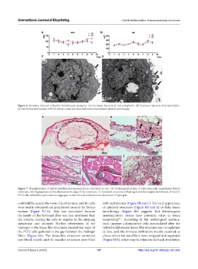
Figure 7. Transplantation of hybrid biofabricated neurosecretory structures in vivo. (A) Pathological section of subcutaneously transplanted hybrid
constructs. (B) Angiogenesis can be observed at the edge of the constructs. (C) Sandwich structures of hydrogels and electrospun membranes. (D and E)
PC12 cells in biofabricated constructs aggregate in nanofiber membranes in the interstices of hydrogels.
could still be seen in the tissue-like structures, and the cells with erythrocytes (Figure 8B and C). The local appearance
were mostly colonized and proliferated around the fibrous of spheroid structures (Figure 8D and E) or flaky tissue
septum (Figure 7C–E). This was speculated because morphology (Figure 8F) suggests that heterozygous
the inside of the hydrogel fiber was less nutritious than neurosecretory tissues have potential value in tissue
the outside, causing the cells to migrate to the spinning remodeling . According to the pathological sections,
[41]
membrane and colonize. Further observation of the local immune inflammatory cells accumulated after the
hydrogel in the tissue-like structures showed that most of hybrid biofabricated tissue-like structure was transplanted
the PC12 cells gathered in the gap between the hydrogel in vivo, and the immune infiltration mainly occurred at
fibers (Figure 8A). The tissue-like structures contained places where the nanofibers were wrapped and separated
new blood vessels, and the vascular structures were filled (Figure S3A), which may be related to the local stimulation
Volume 9 Issue 2 (2023) 137 https://doi.org/10.18063/ijb.v9i2.659