
Page 146 - IJB-9-2
P. 146
International Journal of Bioprinting Hybrid biofabrication of neurosecretory structures
A B C
D E F
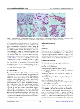
Figure 8. Tissue remodeling of neurosecretory organoids. (A) Hybrid fabrication of neurosecretory structures transplanted in vivo for 14 days.
(B and C) Angiogenesis and lamellar organoid-like structure formation. (D–F) Spherical and sheet-like remodeling tissue structures.
of the nanofibers as foreign bodies. In the selection of Acknowledgments
raw materials for electrospinning, PLLA and gelatin with
good biocompatibility are preferred, both of which are None.
FDA-approved biodegradable polymer materials for in Funding
vivo transplantation and have been proven to have good
histocompatibility. When the pathological sections were This study was supported by the Anhui Provincial Natural
examined at high magnification, immune infiltration was Science Foundation (2208085MH251), the Anhui Medical
not obvious in the local areas where the biofabricated University Scientific Research Fund (No. 2021xkj131), and
structures were reconstructed to form a tissue-like body the Basic and Clinical Cooperative Research and Promotion
(Figure S3B–D). Therefore, we hypothesize that during Program of Anhui Medical University (2022xkjT024).
the early stage of tissue remodeling, immune infiltration
may be involved in the process of tissue remodeling Conflict of interest
and angiogenesis. We speculate that the neurosecretory The authors declare no competing financial interest.
structures produced by hybrid biofabrication can self-
assemble in vivo for tissue remodeling and construct Author contributions
functional vascularized structures.
Conceptualization: Hongwei Cheng, Qing Lan
4. Conclusion Formal analysis: Xingliang Dai, Xuefeng Tian, Shengcai
Gu, Peng Gao
The neurosecretory tissue-like structures were prepared by Investigation: Xingliang Dai, Xuefeng Tian, Shengcai Gu,
combining extrusion 3D bioprinting and electrospinning Peng Gao
technology in vitro, using sodium alginate/gelatin/ Methodology: Xingliang Dai, Xuefeng Tian, Shengcai Gu,
fibrinogen as the matrix composition and supporting Peng Gao
structure. The overall mechanical strength and structural
stability of the hydrogel scaffolds were increased by the Supervision: Hongwei Cheng, Qing Lan
superimposition of PLLA/gelatin electrospun nanofibers. Writing – original draft: Xingliang Dai, Xuefeng Tian, Yafei
The results showed that the neurosecretory cells had Yang, Huaixu Li, Peng Gao
high activity and maintained their original secretory Writing – review and editing: Hongwei Cheng, Qing Lan
function in vitro and showed high growth activity, Ethics approval and consent to participate
self-assembly vascularization, and tissue remodeling
potential after transplantation in vivo. This new method The authors are accountable for all aspects of the work in
allows for the biological manufacture of neurosecretory ensuring that questions related to the accuracy or integrity
structures in vitro as well as provides a new engineering of any part of the work are appropriately investigated and
manufacturing platform for hormone substitution research resolved. All animal experiments were conducted under
of neurosecretory structures. the supervision of the Animal Research Ethics Committee
Volume 9 Issue 2 (2023) 138 https://doi.org/10.18063/ijb.v9i2.659