
Page 144 - IJB-9-2
P. 144
International Journal of Bioprinting Hybrid biofabrication of neurosecretory structures
3.3. Hybrid biofabricated neurosecretory structures the cells maintained good activity and secretory function
maintain biological characteristics and secretion (Figure 6A and B). Moreover, TEM showed the presence
To compare the biological characteristics of neurosecretory of efflux vesicles in the cytoplasm and membrane of
cells after hybrid biofabrication, the expression of PC12- PC12 cells (Figure 6C and D), demonstrating the function
specific proteins MAP2 and tubulin-β was detected by of MEK and NE efflux from hybrid PC12 cells through
Western blotting. The expression of MAP2 and tubulin-β vesicles.
in the PLLA/gelatin membrane-implantation group, 3.4. Angiogenesis and tissue remodeling of hybrid
hydrogel-mixed cell group, and hybrid biofabrication biofabricated neuroendocrine structures in vivo
group was significantly higher than that in the 2D
group (Figure 5A and B). The results suggested that the Hybrid biofabrication was used to produce neurosecretory
PC12 cells in the hybrid biofabrication group maintained structures for transplantation in nude mice to evaluate the
their biomarkers and biological characteristics. stability in vivo and tissue remodeling potential to form
Neurosecretory PC12 cells have typical secretory neurosecretory structures. The mice were sacrificed 3 weeks
characteristics, including the secretion of MEK and NE after subcutaneous embedding transplantation, and grafts
during in vitro culture [36,37] . ELISA was used to detect the were taken for pathological staining (Figure 7A). Tissue-
amount of MEK and NE secreted by PC12 cells after hybrid like morphology was formed on the surface of the hybrid
biofabrication and 3D bioprinting. Similar to the 2D biofabricated structures, alongside evidence of angiogenesis
culture group, the PC12 cells in the hybrid biofabrication (Figure 7B). The emerging new blood vessels could provide
group could secrete MEK and NE stably throughout nutrients and oxygen for the tissue structures, which is the
the entire culture cycle. During the 9-day follow-up, the premise of tissue remodeling and regeneration, suggesting
secretion of MEK and NE remained stable in the hybrid that heterozygous structures can be potentially used to
biofabrication group and 2D culture group, suggesting that reconstruct tissue structures. Electrospinning separation
A B
C
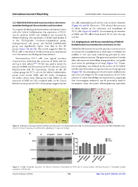
Figure 4. Pathological staining of hybrid biofabricated structures. (A) Hematoxylin and eosin (H and E) staining of structures after hybrid biofabrication.
(B and C) H and E staining of scaffolds after culturing for 7 days and 14 days.
A B
Figure 5. Cellular biomarker detection. (A) Western blotting of biomarkers in PC12 cells in various models. (B) Quantification of biomarker protein
expression in PC12 cells.
Volume 9 Issue 2 (2023) 136 https://doi.org/10.18063/ijb.v9i2.659