
Page 143 - IJB-9-2
P. 143
International Journal of Bioprinting Hybrid biofabrication of neurosecretory structures
membrane. Next, the mechanical compression moduli of tissue remodeling and better simulates the structure and
the hybrid manufactured structure and simple bioprinted morphology of organs [40-44] .
hydrogel structure were tested. The results showed that the
compression moduli of the hybrid manufacturing structure 3.2. Hybrid biofabrication maintains cell viability
and bioprinted hydrogel structure was 49.93±1.79 kPa and and secretory function of neurosecretory structure
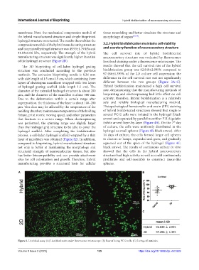
41.83±6.06 kPa, respectively. The strength of the hybrid The cell survival rate of hybrid biofabricated
manufacturing structure was significantly higher than that neurosecretory structure was evaluated by fluorescence
of the hydrogel structure (Figure 2D). live/dead staining under a fluorescence microscope. The
The 3D bioprinting of cell-laden hydrogel grating results showed that the cell survival rate of the hybrid
structure was conducted according to the previous biofabrication group was 92.849±2.995% compared to
methods. The extrusion bioprinting nozzle is 0.26 mm 97.486±1.395% of the 2D culture cell suspension; the
with side length of 1.5 cm×1.5 cm, which containing three difference in the cell survival rate was not significantly
layers of electrospun nanofibers wrapped with two layers different between the two groups (Figure 3A–C).
of hydrogel grating scaffold (side length 1.5 cm). The Hybrid biofabrication maintained a high cell survival
diameter of the extruded hydrogel structure is about 200 rate, demonstrating that the manufacturing methods of
μm, and the diameter of the nanofiber is about 500 nm. bioprinting and electrospinning had little effect on cell
Due to the deformation within a certain range after activity; therefore, hybrid biofabrication is a relatively
superposition, the thickness of the layer is about 100–200 safe and reliable biological manufacturing method.
μm. This data may be affected by the temperature of the Histopathological hematoxylin and eosin (HE) staining
molding chamber, maintenance temperature of the holding of hybrid biofabricated structures showed that single or
fixture, print nozzle moving speed, and other parameters several PC12 cells were isolated in the hydrogel (black
that fluctuate in a certain range. When electrospinning arrow) and separated by parallel nanofiber PLLA/gelatin
th
was performed, the spinning range was slightly larger (white arrow) layer-by-layer (Figure 4A). On the 7 day
than the hydrogel grid structure to be able to cover the of culture, the cells were uniformly distributed in the
hydrogel scaffold. After completing the biofabrication hydrogel as small spheres (Figure 4B, black arrow). After
process, a cell-laden hydrogel scaffold wrapped by a thin 14 days of culture, the cells formed larger cell spheres
layer of nanofibers was obtained (Figure S2). In addition, in clusters or lumps, expanded and grew, and gradually
compared to bioprinting, hybrid manufactured structure squeezed out of the space of the hydrogel (Figure 4C,
not only is better at maintaining the morphology and black arrow). The results of continuous culture in vitro
structural strength of neuroendocrine tissues, but also showed that the cells in the hybrid neurosecretory
has better biocompatibility and can provide attachment structure had high activity as well as could continuously
sites for cell colonization and growth. Therefore, hybrid proliferate and self-assemble to construct tissue-like
manufacturing provides a structural basis for cellular spheres.
A B
C
Figure 3. Live/dead assay. (A) Live/dead stain under fluorescence microscope. (B) Rate of living PC12 cells. (C) Living cell statistics.
Volume 9 Issue 2 (2023) 135 https://doi.org/10.18063/ijb.v9i2.659