
Page 217 - IJB-9-2
P. 217
International Journal of Bioprinting Three-dimensional bioprinting in toxicological research
which consists of encapsulated human induced pluripotent 9. Clinical use
stem cell (hiPSC)-originated hepatocytes, HUVECs and
adipose-derived stem cells (ADSCs), was printed in a pattern Non-biological and biological liver support is available for
that mimics the liver lobule structure. They used 1%, 2.5%, the treatment of patients with acute liver failure. Biological
and 5% (w/v) GelMA to encapsulate endothelial cells and methods take advantage of the functional capacity of
mesenchymal stem cells. After printing, they maintained xenogeneic or human-derived liver cells, thus supporting
the tri-culture and observed the expression of fetal hepatic the function of the patient’s liver. These functions include
marker α-fetoprotein, albumin (ALB), hepatocyte nuclear detoxification, metabolic functions, and synthesis of proteins
factor 4α (HNF4α), and transthyretin. This model showed and other molecules. One of the most effective clinically
the expression of different CYP450 enzymes too, such as used bioartificial liver devices is the AMC-Bio-Artificial
the, CYP2B6, CYP2C9, and CYP2C19, and on the addition liver (AMC-BAL) system, a product developed by a research
of rifampicin, the CYP3A4, CYP2C9, and CYP2C19 were group in Netherlands. This product is a hollow fiber,
induced [133] . Faulkner-Jones et al. developed a 3D model polysulfone-coated bioreactor and plasmapheresis system.
10
by valve-based inkjet bioprinting [134] . They printed hiPSCs At least 1 × 10 viable human (previously porcine) liver cells
and human embryonic stem cells and the cells were in a 3D configuration are attached to a nonwoven material
differentiated into hepatocyte-like cells. Differentiated cells in a hydrophilic polyester matrix. The matrix is 4 mm thick
2
expressed HNF4α and albumin so this model is suitable for and its total surface is 5610 cm , which are helically wound
drug testing, and the bioprinting process did not affect the around a huge core. Between the layers of the matrix, the
viability and pluripotency of the cells [88,134] . Lei and Wang on-site gas exchange takes place through hollow fibers in a
created a model using ADSCs and primary hepatocytes longitudinal direction. During the treatment, blood of the
to form a complex mini organ with vascular systems [135] . patient is subjected to plasma filtering; the filtered plasma is
With this four-nozzle low-temperature technique, the received by the bioreactor that perfuses the blood cells. One
printing of liver organoid and other complex tissue can be of the most important qualities of AMC-BAL is the direct
performed (Table 2) [88,135] . relationship between the small islets of liver cells and the
incoming plasma, and its structure ensures optimal mass for
Due to the complexity and coordinated functioning liver cell transfer and direct oxygen supply [21,136-140] .
of human organs, 3D printing faces an extremely difficult
challenge. In recent years, research has proven that we are 3D tissue printing may be particularly suitable for the
getting closer to printing artificial tissues that function regeneration and/or replacement of diseased or damaged
largely similar to the original organ. As soon as it becomes tissues. In such a case, it is important to design a proper
possible to print tissues that are identical in structure and structure so that the cells can have the correct polarity and
function, the fields of toxicology, personalized medicine, function. When using non-synthetic scaffolds, decellularized
and regenerative medicine will usher in the era of liver tissue is considered an extracellular matrix. The
tremendous development. Despite the many difficulties technique involves decellularizing the target organ and
in the printing of artificial 3D tissues, it has been proven removing all living cells and debris to leave behind the intact
that 3D printed tissues could ensure fast and efficient drug extracellular skeleton. The quality of the matrix is then
testing in the future [82,93] . checked and recellularized with healthy, tissue-specific cells.
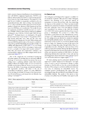
Table 2. Tissue engineered liver models for drug testing or clinical use
Cell type Bioink Results
Hepatocytes Gelatin Hepatocytes showed high viability for more than 2 months
and their biological function remained intact
Primary human hepatocytes, hepatic stellates, NovoGelR 2.0 hydrogel The cells were viable for 28 days
HUVEC cells, and non-parenchymal cells
Primary mouse hepatocytes Galactosylated alginate The viability was >85% after 2 days
HUVEC - Multi-layered model for testing hepatotoxicity
Primary mouse hepatocytes Alginate The cells were viable for 14 days
HepG2, BM-MSCs Decellularized extracellular matrix Liver tissue model
hiPSCs, hESCs RC-6 and alginate The viability of cells decreased to >55% after 1 day
Primary hepatocytes - The cells were viable for 60 days
HUVEC, Human umbilical vein endothelial cells; BM-MSCs, Bone marrow mesenchymal stem cells; hiPSCs, human induced pluripotent stem cell;
hESCs, human embryonic stem cells.
Volume 9 Issue 2 (2023) 209 https://doi.org/10.18063/ijb.v9i2.663