
Page 219 - IJB-9-2
P. 219
International Journal of Bioprinting Three-dimensional bioprinting in toxicological research
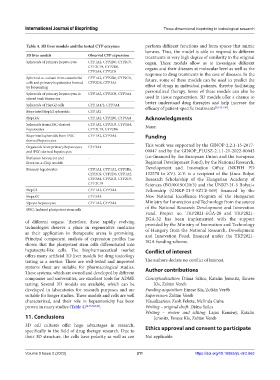
Table 4. 3D liver models and the tested CYP enzymes perform different functions and form spaces that mimic
lumens. Thus, the model is able to respond to different
3D liver models Observed CYP expression treatments in very high degree of similarity to the original
Spheroids of primary hepatocytes CYP1A2, CYP2B6, CYP2C9, organ. These models allow us to investigate different
CYP2C19, CYP2D6, organs and their diseases at molecular level as well as the
CYP3A4, CYP2C8 response to drug treatments in the case of diseases. In the
Spheroid co-culture from endothelial CYP1A2, CYP2B6, CYP2C9, future, some of these models can be used to predict the
cells and primary hepatocytes formed CYP2D6, CYP3A4
by bioprinting effect of drugs in individual patients, thereby facilitating
Spheroids of primary hepatocytes in CYP1A2, CYP2C9, CYP3A4 personalized therapy. Some of these models can also be
stirred tank bioreactor used in tissue regeneration. 3D models offer a chance to
better understand drug therapies and help increase the
Spheroids of HepG2 cells CYP1A1/2, CYP3A4
efficacy of patient-specific treatments [8,142-146] .
Bioprinted HepG2 spheroids CYP1A2
HepaRG CYP1A2, CYP2B6, CYP3A4 Acknowledgments
Spheroids from iPSC-derived CYP1A2, CYP2C9, CYP3A4, None.
hepatocytes CYP2C19, CYP2D6
Bioprinted spheroids from iPSC CYP1A2, CYP3A4 Funding
derived hepatocytes
Organoids from primary hepatocytes CYP3A4 This work was supported by the GINOP-2.2.1-15-2017-
and iPSC-derived hepatocytes 00047 and by the GINOP_PLUSZ-2.1.1-21-2022-00043
Perfusion bioreactor and - (co-financed by the European Union and the European
Liver-on-a-Chip models Regional Development Fund), by the National Research,
Primary hepatocytes CYP1A1, CYP1A2, CYP2B6, Development and Innovation Office (NKFIH PD
CYP2C9, CYP2D6, CYP1A2, 132570 to ZV). Z.V. is a recipient of the János Bolyai
CYP3A4, CYP2C8, CYP2C9, Research Scholarship of the Hungarian Academy of
CYP2C19 Sciences (BO/00190/20/5) and the ÚNKP-21-5 Bolyai+
HepG2 CYP1A2, CYP3A4 Fellowship (ÚNKP-21-5-SZTE-169) financed by the
HepaRG CYP3A4 New National Excellence Program of the Hungarian
Upcyte hepatocytes CYP1A2, CYP3A4 Ministry for Innovation and Technology from the source
iPSC: Induced pluripotent stem cells of the National Research Development and Innovation
Fund. Project no. TKP2021-EGA-28 and TKP2021-
EGA-32 has been implemented with the support
of different organs. Therefore, these rapidly evolving provided by the Ministry of Innovation and Technology
technologies deserve a place in regenerative medicine of Hungary from the National Research, Development
as their application in therapeutic arena is promising. and Innovation Fund, financed under the TKP2021-
Principal component analysis of expression profiles has EGA funding scheme.
shown that the pluripotent stem cells differentiated into
hepatocyte-like cells. The biopharmaceutical market Conflict of interest
offers many artificial 3D liver models for drug toxicology
testing as a service. These are well-tested and improved The authors declare no conflict of interest.
systems theat are suitable for pharmacological studies. Author contributions
These systems, which are owned and developed by different
companies and universities, are excellent tools for ADME Conceptualization: Diána Szűcs, Katalin Jemnitz, Emese
testing. Several 3D models are available, which can be Kis, Zoltán Veréb
developed in laboratories for research purposes and are Funding acquisition: Emese Kis, Zoltán Veréb
suitable for longer studies. These models and cells are well Supervision: Zoltán Veréb
characterized, and their role in hepatotoxicity has been Visualization: Zsolt Fekete, Melinda Guba
proven in many studies (Table 4) [4,35,38,141] . Writing – original draft: Diána Szűcs
Writing – review and editing: Lajos Kemény, Katalin
11. Conclusions Jemnitz, Emese Kis, Zoltán Veréb
3D cell cultures offer huge advantages in research,
specifically in the field of drug therapy research. Due to Ethics approval and consent to participate
their 3D structure, the cells have polarity as well as can Not applicable.
Volume 9 Issue 2 (2023) 211 https://doi.org/10.18063/ijb.v9i2.663