
Page 241 - IJB-9-2
P. 241
International Journal of Bioprinting Coronary and peripheral artery disease. State of the art.
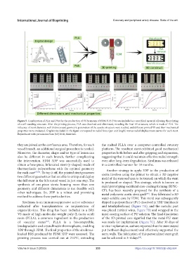
Figure 5. Combination of PLA and PVA for the production of VS by means of FDM 3DP. PVA was included as a sacrificial material allowing the printing
of a self-standing structure. After the printing process, PVA was dissolved and eliminated, revealing the final VS structure, which is made of PLA. The
influence of stent diameter, wall thickness and geometric parameters of the auxetic structure were studied, and different printed VS and their mechanical
properties were evaluated. Graphics included in the figure correspond to radial force (per unit length) versus radial displacement curves for each stent.
Reproduced with permission from [62] 2018, Materials.
they are joined at the confluence area. Therefore, for each the melted PLLA over a computer-controlled rotatory
vessel branch, an additional surgical procedure is needed. platform. The resultant stent exhibited good mechanical
Moreover, the diameter, shape and/or type of lesion can properties both before and after gripping and expansion,
also be different in each branch, further complicating suggesting that it could maintain effective radial strength
the intervention. FDM 3DP was successfully used to even after long-term degradation. Sirolimus was released
obtain a fone-piece, bifurcated stent (y-shaped) made of in a controlled manner for 18 months.
thermoplastic polyurethane with the optimal geometry Another strategy to apply 3DP to the production of
for each case [65,66] . To top it off, the printed stent possesses stents involves using the printer to obtain a 3D negative
two different geometries that are able to crimp and deploy mold of the stenosed area to be treated, on which the stent
the full stent in the bifurcated vessel in just one step. The is produced or shaped. This strategy, which is known as
synthesis of one-piece stents housing more than one rapid prototyping sacrificial core-coating forming (RPSC-
geometry and different dimensions is not feasible with CF), has been recently proposed for the synthesis of a
other techniques. So, 3DP is a robust and promising metal-polymeric aortic stent graft . They fabricated a 3D
[68]
method to address these particularities.
water-soluble core by FDM. This mold was subsequently
Sirolimus is an immunosuppressive active substance dipped in polyurethane (PU) dissolved in THF (imidazole
indicated after transplantation or implantation of and tetrahydrofuran) (Figure 7A), and the metallic part
organs/devices. This drug has been included in a BRS was placed (nitinol wires, Figure 7B) and wrapped in the
VS made of high-molecular-weight poly (L-lactic acid) inner coating surface of PU solution. The final dissolution
resin (PLLA), a common ingredient in the production of the 3D-printed core signified that the metal-PU stent
of vascular stents [67] . PLLA is a biocompatible, was ready for implantation (Figure 7C). After 30 days of
biodegradable and crystallizable thermoplastic, enabling in vivo implantation, authors reported that the stent stayed
3DP through FDM. The final properties of the sirolimus- put (without displacement) and effectively supported the
loaded BRS produced by FDM 3DP were assessed. The aorta walls. The fabrication of this personalized stent graft
printing process was carried out at 210°C, extruding can be achieved in 3–4 days .
[68]
Volume 9 Issue 2 (2023) 233 https://doi.org/10.18063/ijb.v9i2.664