
Page 243 - IJB-9-2
P. 243
International Journal of Bioprinting Coronary and peripheral artery disease. State of the art.
Figure 7. (A) Pulling platform for deposition process over the sacrificial mold obtained by means of 3DP. (B) Shaped nitinol wires to be used in the final
stent. (C) Final aspect of aortic metal-PU stents with different shapes (branched and straight). Reproduced with permission from [68] 2020, Medical
Engineering and Physics.
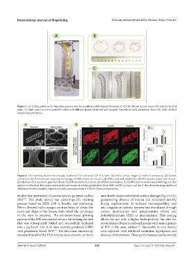
Figure 8. (A) Scanning electron microscopy of printed PLA stents: (a) full PLA stent; (b) entire surface image; (c) exterior connection; (d) interior
connection. (B) Confocal laser scanning microscopy (CLSM) of smooth muscle cells (SMC, red) and endothelial cells (EC, green) seeded over the pro-
duced stents. PLA stands for pure PLA stents; PLADP stands for PLA stents after PEI immobilization; PLADPH refers to stents loaded with heparin after
surface modification. Bar charts represent the percentage of cellular proliferation (both SMC and EC) at day 1 and day 3, thus demonstrating significant
differences between samples. Reproduced with permission from [14] 2019, Chemical Engineering.
enabled the production of patient-specific polymer-carbon and clearly connected without surface damage (Figure 8A),
BRS . This study proves that patient-specific stenting guaranteeing absence of trauma and structural stability
[70]
process based on MEX 3DP is feasible and promising. during implantation. To enhance biocompatibility and
Fibrin-directed radio-opaque contrast helps to obtain the anti-coagulation activity, heparin was introduced through
mold and shape of the lesion, from which the 3D design surface modification with polydopamine (PDA) and
of the stent is prepared. The extrusion-based printing polyethyleneimine (PEI) as intermediates. This coating
process of the BRS was carried out as a flat rectangular slab allows for not only a higher hydrophilicity, but also the
that was subsequently folded and successfully deployed crosslinking of heparin carboxyl groups with amino groups
into a pig heart. Lee et al. have recently produced a BRS of PEI in the stent surface . Successful in vivo studies
[14]
with pneumatic-based 3DP . The electronic microscopy were reported, with inhibited neointima hyperplasia and
[14]
revealed that all of the PLA strands were smooth, uniform absence of thrombosis. These performances can be entirely
Volume 9 Issue 2 (2023) 235 https://doi.org/10.18063/ijb.v9i2.664