Page 380 - IJB-9-2
P. 380

International Journal of Bioprinting  3D gel-printed β-TCP/TiO2 porous scaffolds for cancellous bone tissue engineering


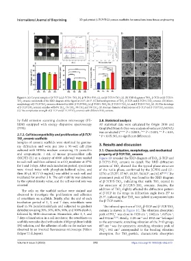
            Figure 1. (A) Ceramic samples of β-TCP (a), β-TCP/1-TiO  (b), β-TCP/3-TiO  (c), and β-TCP/5-TiO  (d). (B) XRD diagram of TiO , β-TCP, and β-TCP/5-
                                                                            2
                                                                                               2
                                                 2
                                                             2
            TiO  ceramic and details of the XRD diagram of the figure at 24.5°–26.5°. (C) Infrared spectrum of TiO , β-TCP, and β-TCP/5-TiO  ceramic. (D) Micro-
                                                                              2
                                                                                                2
              2
            morphology of β-TCP/TiO  ceramics detected by SEM: β-TCP/TiO  (a), β-TCP/1-TiO  (b), β-TCP/3-TiO  (c), and β-TCP/5-TiO  (d). (E) The shrinkage
                                                                                              2
                             2
                                                                               2
                                                                   2
                                                       2
            of β-TCP/TiO  ceramic samples with 0% TiO , 1% TiO , 3% TiO  and 5% TiO . (F) Average diameter of micropores of β-TCP and β-TCP/TiO  ceramics.
                                               2
                                                              2
                                                     2
                                         2
                                                                                                      2
                     2
            (G) The compressive strength of β-TCP and β-TCP/TiO ceramics with different TiO  content.
                                                                  2
                                               2
            by field emission scanning electron microscopy (FE-  2.8. Statistical analysis
            SEM) equipped with energy dispersive spectroscopy   All statistical data were calculated by Origin 2018 and
            (EDS).                                             GraphPad Prism 8. One-way analysis of variance (ANOVA)
                                                               was conducted (**** P < 0.0001; *** P < 0.001; ** P < 0.01;
            2.7.2. Cell biocompatibility and proliferation of β-TCP/  * P < 0.05; NS, no significant difference).
            TiO  ceramic scaffolds
               2
            Samples of ceramic scaffolds were sterilized by gamma-
            ray  diffraction  and  were  put  into  a  96-well  cell  plate   3. Results and discussion
            cultured with MEMα medium containing 1% penicillin   3.1. Characterization, morphology, and mechanical
            and streptomycin. 1 mL of mouse preosteoblast cells   property of β-TCP/TiO  ceramic
                                                                                  2
            (MC3T3-E1) at a density of 6000 cells/well were seeded   Figure 1B revealed the XRD diagram of TiO , β-TCP and
                                                                                                   2
            in each well and then cultured in a CO incubator at 37°C   β-TCP/5-TiO  ceramic in detail. The XRD diffraction
                                           2
                                                                          2
            for 1 and 3 days. After each incubation period, specimens   pattern of TiO  showed that the typical phase structure
                                                                           2
            were rinsed twice with phosphate-buffered saline, and   of the rutile phase, confirmed by the JCPDS card (21-
            then 10 μL MTT (5 mg/mL) was added to each well and   1276) at 25.25°, 37.96°, 48.20°, 54.143°, and 62.87° . The
                                                                                                       [35]
            incubated for another 3 h. The cell viability was detected   prominent peak of TiO  was found in the XRD diagram
                                                                                  2
            by the optical density value, and the cell survival rate was   of β-TCP/5-TiO , indicating that rutile TiO  existed in
                                                                                                    2
                                                                            2
            counted.                                           the structure of β-TCP/5-TiO  ceramic. Besides, the
                                                                                         2
               The cells on the scaffold surface were stained and   addition of TiO  slightly affected the diffraction pattern
                                                                            2
            observed to investigate the proliferation and adhesion   of β-TCP in the range in diffraction angle from 25° to
            of osteoblasts on scaffolds. Briefly, after the end of each   25.5°, indicating that TiO  was indeed incorporated into
                                                                                    2
            incubation period of 1, 3,  and 7 days, osteoblasts were   the β-TCP matrix.
            fixed in 5% paraformaldehyde and subjected to gradient   The infrared spectrum of TiO , β-TCP and β-TCP/TiO
                                                                                         2
                                                                                                             2
            dehydration using 30%, 50%, 80%, 90%, and 100% alcohol,   ceramic is shown in Figure 1C. The vibration absorption
            followed by SEM observation. Meanwhile, after 1, 3, and   peak of PO  was close to 1120 cm , 1042cm , 607cm ,
                                                                                                            -1
                                                                                                    -1
                                                                                           -1
                                                                         3-
                                                                        4
            7 days of incubation in a cell incubator, the osteoblasts on   and 552cm -1[36] . Briefly, 1120 cm and 1042 cm belonged
                                                                                         -1
                                                                                                    -1
            scaffolds were also dyed with calcein AM/propidium iodide   to the asymmetric stretching vibration of the P=O group;
            (PI) solution, and the adhesion of cells on the surface was   607 cm  was the symmetric stretching vibration peak of
                                                                     -1
            observed by an inverted fluorescence microscope (Nikon   PO ; 552 cm corresponded to the bending vibration
                                                                           -1
                                                                  3-
                                                                 4
            Eclipse Ti-E, Japan).                              absorption. For TiO particle, characteristic absorption
                                                                                2
            Volume 9 Issue 2 (2023)                        372                     https://doi.org/10.18063/ijb.v9i2.673
   375   376   377   378   379   380   381   382   383   384   385