
Page 385 - IJB-9-2
P. 385
International Journal of Bioprinting 3D gel-printed β-TCP/TiO2 porous scaffolds for cancellous bone tissue engineering
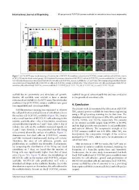
Figure 7. (A) The MTT assay results showing cell viability rate of MC3T3-E1 osteoblasts cultured on β-TCP/TiO ceramic scaffolds with different content
2
of TiO (CK was the blank control group). (B) Osteoblast fluorescence detection of MC3T3-E1 cells on β-TCP/TiO ceramic scaffolds for 1, 3, and 7 days.
2
2
(C) Osteoblast fluorescence detection of dead MC3T3-E1 cells on β-TCP/TiO ceramic scaffolds for 1, 3, and 7 days. (D) Corresponding osteoblast fluores-
2
cence detection of living/dead cells of MC3T3-E1 cells on β-TCP/TiO ceramic scaffolds after 7 days of incubation. (E) Cell morphologies of MC3T3-E1
2
osteoblast cells cultured on β-TCP/TiO ceramic scaffolds: β-TCP/TiO (a), β-TCP/1-TiO (b), β-TCP/3-TiO (c), and β-TCP/5-TiO (d).
2 2 2 2 2
scaffold has no cytotoxicity and stimulates cell growth. scaffold has good cytocompatibility and was conducive
Besides, all scaffolds were verified to have a similar to the growth of osteoblast cells.
outcome of cell viability in the MTT assay. The results also
confirmed that β-TCP/TiO ceramic scaffolds have good 4. Conclusion
2
biocompatibility and cytocompatibility.
Cell fluorescence staining was examined to observe The present work demonstrated the fabrication of β-TCP/
the cell proliferation and adhesion of osteoblast cells on TiO ceramic porous scaffolds for bone tissue engineering
2
the surface of β-TCP/TiO scaffolds (Figure 7B). Despite using a 3D gel-printing sintering for the first time. The
2
only a small number of MC3T3-E1 cells adhering to the shrinkage ratio with filling rates of 20%, 30%, and 40% was
ceramic scaffolds after 1 day of incubation, osteoblasts 56.51%, 54.96%, and 53.57%, respectively. The porosity
cells proliferated rapidly on 3 and 7 days. After 1 day of of the sintered scaffolds ranged from 65.55% to 66.39%,
culture, the cells on the scaffolds increased rapidly on and the high porosity was conducive to cell proliferation
3 and 7 days. Notably, it was perceived that the living and nutrient delivery. The compressive strength of the
cells covered almost the surface of scaffolds. Figure 7C β-TCP ceramics scaffold was 0.35 MPa. After TiO was
2
demonstrates that dead cells on β-TCP/TiO ceramic incorporated, the compressive strength of the ceramics
2
scaffolds increased simultaneously during cell culture. increased to 0.72 MPa, which meets the requirements of
Therefore, the state of cell adhesion, growth, and cancellous bone.
proliferation on scaffolds was favorable. Furthermore, After immersion in SBF for weeks, the Ca/P ratio on
by comparing the distribution of the living and dead the surface of ceramics scaffolds increased, implying the
osteoblasts on 7 days, we found that the number of formation of a bone-like apatite layer and bioactivity.
living cells was significantly higher than that of dead Furthermore, the in vitro study demonstrated that both
cells (Figure 7D). Figure 7E shows the morphology of the β-TCP scaffold and β-TCP/TiO porous scaffolds were
2
MC3T3-E1 cells on the surface of β-TCP/TiO ceramic favorable for cell adhesion, growth and proliferation of
2
scaffolds. MC3T3-E1 cells proliferated well with a MC3T3-E1 osteoblast cells. In conclusion, the β-TCP/TiO 2
stretched cell shape on the scaffolds. Besides, due to the porous scaffold has a lot of potential to be a promising
abundant micropores on the surface, the cells adhered to bone repair scaffold in craniomaxillofacial and orthopedics
the scaffolds firmly. To sum up, the β-TCP/TiO ceramic surgery.
2
Volume 9 Issue 2 (2023) 377 https://doi.org/10.18063/ijb.v9i2.673