
Page 382 - IJB-9-2
P. 382
International Journal of Bioprinting 3D gel-printed β-TCP/TiO2 porous scaffolds for cancellous bone tissue engineering
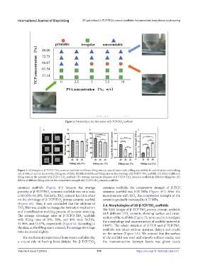
Figure 2. Printability in the fabrication of β-TCP/TiO scaffolds
2
Figure 3. (A) Samples of β-TCP/3-TiO ceramics scaffolds in different filling rates (a1 and a2: stents with a filling rate of 40%; b1 and b2: stents with a filling
2
rate of 30%; c1 and c2: stents with a filling rate of 20%). (B) Effect of different filling rates on the shrinkage of β-TCP/3-TiO scaffolds. (C) Effect of different
2
filling rates on the porosity of β-TCP/3-TiO scaffolds. (D) Average macropore diameter of β-TCP/3-TiO ceramics scaffolds in different filling rate. (E)
2
2
Effects of different filling rates on the compressive strength of β-TCP/3-TiO ceramics scaffolds.
2
ceramics scaffolds (Figure 4D) because the average ceramics scaffolds, the compressive strength of β-TCP
porosity of β-TCP/TiO ceramics scaffolds was on a scale ceramics scaffold was 0.35 MPa (Figure 4C). After the
2
of 65.55%–66.39%. Similarly, TiO content has little effect incorporation with TiO , the compressive strength of the
2
2
on the shrinkage of β-TCP/TiO porous ceramic scaffold ceramics gradually increased to 0.72 MPa.
2
(Figure 4E). Thus, it was concluded that the addition of 3.4. Morphologies of 3D β-TCP/TiO scaffolds
TiO filler was unable to change the formation mechanism The SEM images of β-TCP/TiO porous ceramic scaffolds
2
2
and crystallization-molding process of ceramic sintering. with different TiO contents, showing surface and cross-
2
The average shrinkage rates of β-TCP/3-TiO scaffolds section of the scaffolds (Figure 5), were used to investigate
2
2
with filling rates of 20%, 30%, and 40% were 56.51%, the morphology and microstructure of scaffold sintered at
54.96%, and 53.57%, respectively (Figure 6). According to 1100°C. The whole structure of β-TCP and β-TCP/TiO
the data, as the filling rate increased, the average shrinkage scaffolds was intact without apparent defects and cracks
2
rates decreased slightly.
on the surface (Figure 5A). We noticed that the surface
The mechanical properties of bone repair scaffolds play of the scaffold was even and smooth without cracks, and
a crucial role in healing bone defects. For β-TCP/TiO the interconnection between layers was glossy (scale
2
Volume 9 Issue 2 (2023) 374 https://doi.org/10.18063/ijb.v9i2.673