
Page 65 - IJB-9-2
P. 65
International Journal of Bioprinting Bioprinting in wound dressing and healing
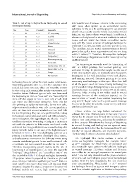
Table 3. List of top 14 keywords for bioprinting in wound interfacial tension. It reduces irritation to the surrounding
dressing and healing skin tissue when applied as an extracellular matrix
substitute for the skin. Its swelling property can promptly
No. Freq Centrality Keywords absorb tissue exudate, keep the wound clean, reduce wound
1 44 0.14 3D bioprinting infection, and thus accelerate wound repair. In addition, it
2 42 0.23 Scaffold shows excellent physical or structural similarity to natural
3 41 0.21 In vitro tissues and can mimic the natural extracellular matrix
4 33 0.12 Wound healing very well. The porous structure of hydrogels facilitates the
5 28 0.14 Tissue transport of oxygen, nutrients, and some growth factors.
6 28 0.17 Stem cell This provides a suitable spatial microenvironment for cell
7 27 0.15 Hydrogel growth during skin tissue regeneration and acts as a drug
delivery pathway . Therefore, biocompatible hydrogels
[55]
8 26 0.12 Drug delivery have a wide range of applications in skin tissue engineering
9 25 0.12 Tissue engineering and biomedicine.
10 23 0.08 Fabrication The technologies currently used for bioprinting of
11 19 0.12 Mesenchymal stem cell skin are inkjet printing, laser-assisted printing, and
12 18 0.05 Extracellular matrix extrusion printing. To gain further insight into the use of
13 16 0.04 Design these printing techniques, we manually identified papers
14 15 0.05 Keratinocyte investigated in this work (excluding review, book chapter,
and meeting abstract). Extrusion printing is the most
technology has so far yielded data from in vitro experiments. commonly used technique in this topic. More than half
Bioprinting-generated skin is a skin-like substitute with of all the papers used extrusion printing technology to
natural skin tissue structure, which can be used to prepare print wound dressings. Inkjet printing is also a commonly
in vitro using cells, extracellular matrix components, and used technology, accounting for about 35% of all papers.
bioactive factors. Different types of cells have been used However, inkjet printing is not widely used in wound
for bioprinting in vitro, so “stem cell” and “mesenchymal dressings because of the restriction imposed on the
stem cell” also appear in Table 3. Stem cells are cells that concentration of bioink. Laser-assisted bioprinting has
can renew and differentiate themselves. Stem cells for only recently begun to be used to print wound dressings
skin printing are epidermal stem cells, dermal stem cells, because of its ability to kill cells. In our survey, only nine
adipose stem cells, melanin stem cells, mesenchymal stem articles used this technique.
cells, etc. Stem cells are an important source of seed cells Cluster analysis can help further understand the
for skin bioprinting. They can solve problems that existing different directions of investigation in this topic. Figure 8
technologies cannot solve, such as the lack of blood vessels, shows that 13 clusters were formed. On the whole, many
sensory receptors, skin appendages, etc. Skardal et al. clusters have overlapping areas, indicating the similarities
[52]
used laser deposition bioprinting techniques to unite stem of their contents. This reflects that the topic of bioprinting
cells with the potential to differentiate into blood vessels in wound dressing and healing has not spawned many
and found that they greatly facilitated wound healing after subdivisions. Table 4 describes the clusters and their ID, size
injury. Growth factor is also one of the high-frequency (number of papers), silhouette, and respective keywords.
keywords in Table 3. The most challenging aspect of skin The following is a short explanation of each cluster:
printing is achieving formation of multiple blood vessels,
and early attempts to overcome this difficulty involve using 3.1. Hydrogel. This cluster contains a series of
vascular endothelial growth factors and keratinized cells works on bioprinted hydrogels. For example, Maver
or scaffolds, but more research is needed in this area . et al. compared the effectiveness of bioprinted and
[56]
[53]
Therefore, incorporating growth factors in the ink of electrostatically spun carboxymethylcellulose hydrogels
bioprinting is an essential part of this topic. The keywords as wound dressings. Diclofenac sodium and lidocaine
“hydrogel,” “drug delivery,” and “extracellular matrix” are were added to the hydrogel preparation for wound pain
also included in Table 3. Hydrogels are highly hydrophilic relief. They not only characterized the physicochemical,
3D polymer networks prepared by chemical or physical structural, and morphological properties of the hydrogels
cross-linking of natural or synthetic polymers that can derived from the two preparations, but also examined
[54]
[57]
swirl rapidly upon water absorption and maintain their their biocompatibility. Abasalizadeh et al. reviewed
structural integrity. The physical properties of the dissolved the hydrogels formed by alginate with inorganic cations
hydrogel include soft texture, high elasticity, and low and their potential application as bioinks in bioprinting.
Volume 9 Issue 2 (2023) 57 http://doi.org/10.18063/ijb.v9i2.653