
Page 145 - IJB-9-3
P. 145
International Journal of Bioprinting Bioprinting tissue-engineered bone-periosteum biphasic complex.
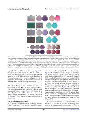
Figure 2. Cell culture and co-culture of rabBMSCs/rabPDSCs. (A) The morphology of rabBMSCs (scale bar = 200 μm). (B) The alkaline phosphatase
(ALP) staining of rabBMSCs after 1 week of osteogenic induction. (C) The Alizarin red S staining of rabBMSCs after 3 weeks of osteogenic differentia-
tion. (D) The oil red O staining of rabBMSCs after 12 days of adipogenic differentiation (scale bar = 50 μm). (E) The Alcian blue staining of rabBMSCs
after 3 weeks of chondrogenic differentiation. (F) The morphology of rabPDSCs (scale bar = 200 μm). (G) The ALP staining of rabPDSCs after 1 week of
osteogenic induction. (H) The Alizarin red S staining of rabPDSCs after 3 weeks of osteogenic differentiation. (I) The oil red O staining of rabPDSCs after
12 days of adipogenic differentiation (scale bar = 50 μm). (J) The Alcian blue staining of rabPDSCs after 3 weeks of chondrogenic differentiation. (K) The
ALP staining of rabBMSCs and rabPDSCs, co-culturing of rabBMSCs and rabPDSCs, as well as their osteogenic induction groups after 1 week. (L) The
Alizarin red S staining of rabBMSCs and rabPDSCs, co-culturing of rabBMSCs and rabPDSCs, as well as their osteogenic induction groups after 3 weeks.
higher than that of 10% mass fraction group (Figure 3B). The storage modulus (G’) of GelMA was shown in the
In addition, the elastic modulus of 5.4 W PLLA/20% HA Figure S2A. It could be seen that under UV irradiation,
group was the highest, which was significantly different the storage modulus (G’) of GelMA increased rapidly,
from that of 5.4 W PLLA/10% HA group. Therefore, in while the dissipation modulus (G’’) was hardly changed.
the subsequent printing process, we adopted the mixed GelMA reached the gelation conversion point after 20 s,
proportion of 5.4 W PLLA with 20% HA to construct the which indicated that the hydrogel needs at least 20 s of UV
bone supporting scaffold of the biphasic complex. irradiation to complete the crosslinking. After 20 s, the G’
Then, we analyzed the biocompatibility of PLLA/ of GelMA continuously improved with the extension of
HA scaffold through CCK-8 experiment and tested the UV irradiation time, indicating that the strength of the
proliferation of rabBMSCs on the 3D-printed scaffolds. hydrogel was getting better. Figure S2B shows the FTIR
The results showed that the number of cells increased spectra of GelMA. There was a characteristic absorption
continuously within 14 days (Figure 3C). Moreover, as peak assigned to peptide bond, in which the amide I
−1
shown by SEM, PLLA/HA scaffold showed staggered appearing in the range 1630–1690 cm is mainly related
porous channels, while rabBMSCs could adhere and stretch to C=O stretching. The degradation curve showed that
on the surface of the scaffold with extended pseudopodia GelMA was rapidly degraded under the action of lysozyme,
(Figure 3D). and it was almost completely degraded on the 14th day
(Figure S2C).
3.3. 3D bioprinting cells pattern In our study, GelMA was used to load rabPDSCs and
In the process of 3D bioprinting, the rheological property rabBMSCs to simulate the periosteum phase and bone
of GelMA plays a key role in forming a stable 3D structure. phase of the biphasic complex, respectively. As shown in
Volume 9 Issue 3 (2023) 137 https://doi.org/10.18063/ijb.698