
Page 354 - IJB-9-3
P. 354
International Journal of Bioprinting Decellularized materials for bioprinting of liver constructs
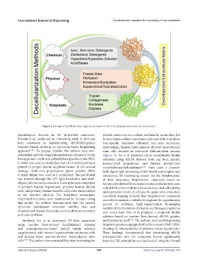
Figure 4. Concept of decellularization approaches based on chemical, physical, and enzymatic procedures.
physiological features in 3D bioprinted constructs. printed constructs in a culture medium for seven days, the
Skardal et al. conducted an interesting study to fabricate human hepatocellular carcinoma cells were able to produce
liver constructs by implementing dECM/HA/gelatin liver-specific functions (albumin and urea secretion).
blended bioink utilizing an extrusion-based bioprinting Interestingly, human bone marrow-derived mesenchymal
approach [124] . To prepare bioinks, the authors used two- stem cells showed an enhanced differentiation process
crosslinker and two-stage polymerization chemistry. In the (Figure 7). Yu et al. prepared photo-crosslinkable bioink
first approach, multi-arm polyethylene glycol acrylate (PEG solutions using dECM derived from pig liver, gelatin,
4-Arm) was used as crosslinker and (4-(2-hydroxyethoxy) methacryloyl prepolymer, and lithium phenyl-2,4,6
phenyl-(2-propyl) ketone as photoinitator. In the second trimethylbenzoylphosphinate [126] . They used a custom-
strategy, multi-arm polyethylene glycol acrylate (PEG built digital light processing (DLP)-based scanningless and
8-Arm) alkyne was used as a crosslinker. The pre-bioink continuous 3D bioprinting system for the biofabrication
was formed through the UV light irradiation and thiol- of liver structures. Biopatterned constructs based on
alkyne polymerization reaction. Liver spheroids comprised hepatocytes derived from human induced pluripotent stem
of primary human hepatocytes, primary human stellate cells (hiPSCs) were cultured for seven days, and cell viability
cells, and primary human Kupffer cells were encapsulated and expression levels of cell-specific genes were evaluated.
in the blended solution. For functional assessment, Live/dead staining showed that biopatterned constructs
bioprinted structures were maintained for 14 days. Using were able to maintain viability throughout the experimental
this model, the authors demonstrated that the printed period. In addition, high-magnification bioimaging
spheroids maintained consistent viability rate, and confirmed the formation of clusters and hexagonal patterns
recapitulated hepatic functions, such as albumin secretion over seven days. Kim et al. prepared a composite bioink
and urea synthesis. solution based on porcine liver-derived dECM, gelatin,
Similarly, Lee et al. developed 3D liver constructs and hyaluronic acid [127] . The authors used multidispensing
using porcine liver-derived decellularized material bioprinting system equipped with a Nano master SMP-III for
and polycaprolactone-based hybrid bioink solution creating the micropatterns of primary mouse hepatocytes.
supplemented with human hepatocellular carcinoma cells Their findings demonstrated that introducing dECM
and human bone marrow-derived mesenchymal stem microparticles into the composite bioink significantly
cells [125] . The authors demonstrated that after incubating the improved 3D printability and mechanical integrity. Overall
Volume 9 Issue 3 (2023) 346 https://doi.org/10.18063/ijb.714