
Page 369 - IJB-9-3
P. 369
International Journal of Bioprinting 3D-printed anistropic meniscus
A B C
D E F
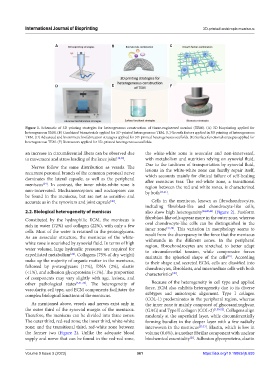
Figure 1. Schematic of 3D printing strategies for heterogeneous construction of tissue-engineered menisci (TEM). (A) 3D bioprinting applied for
heterogeneous TEM. (B) Combined biomaterials applied for 3D-printed heterogeneous TEM. (C) Growth factors applied in 3D printing of heterogeneous
TEM. (D) Advanced and biomimetic biofabrication strategies applied for 3D-printed heterogeneous scaffolds. (E) Surface functional strategies applied for
heterogeneous TEM. (F) Bioreactors applied for 3D-printed heterogeneous scaffolds.
an increase in circumferential fibers can be observed due the white-white zone is avascular and non-innervated,
to movement and stress loading of the knee joint [32,39] . with metabolism and nutrition relying on synovial fluid.
Due to the tardiness of transportation by synovial fluid,
Nerves follow the same distribution as vessels. The
recurrent peroneal branch of the common peroneal nerve lesions in the white-white zone can hardly repair itself,
which accounts mainly for clinical failure of self-healing
dominates the lateral capsule, as well as the peripheral after meniscus tear. The red-white zone, a transitional
meniscus . In contrast, the inner white-white zone is region between the red and white zones, is characterized
[24]
non-innervated. Mechanoreceptors and nociceptors can by both [13,21] .
be found in the meniscus, but are not as sensitive and
accurate as in the synovium and joint capsule . Cells in the meniscus, known as fibrochondrocytes,
[40]
including fibroblast-like and chondrocyte-like cells,
2.2. Biological heterogeneity of meniscus also show high heterogeneity [24,45,46] (Figure 2). Fusiform
Constituted by the hydrophilic ECM, the meniscus is fibroblast-like cells appear more in the outer zone, whereas
rich in water (72%) and collagen (22%), with only a few oval chondrocyte-like cells can be distinguished in the
[47,48]
cells. Most of the water is retained in the proteoglycans. inner zone . This variation in morphology seems to
As an avascular structure, the meniscus of the white- result from the discrepancy in the force that the meniscus
withstands in the different zones. In the peripheral
white zone is nourished by synovial fluid. In terms of high region, fibrochondrocytes are stretched to better adapt
water volume, large hydraulic pressures are required for to circumferential tension, while compressive forces
expeditated metabolism . Collagens (75% of dry weight) maintain the spherical shape of the cells . According
[24]
[49]
make up the majority of organic matter in the meniscus, to their shape and secreted ECM, cells are classified into
followed by proteoglycans (17%), DNA (2%), elastin chondrocytes, fibroblasts, and intermediate cells with both
(<1%), and adhesion glycoproteins (<1%). The proportion characteristics .
[48]
of components may vary slightly with age, lesions, and
other pathological states [5,41-44] . The heterogeneity of Because of the heterogeneity in cell type and applied
vascularity, cell type, and ECM components facilitates the force, ECM also exhibits heterogeneity due to its diverse
complex biological functions of the meniscus. subtypes and anisotropic alignment. Type I collagen
(COL-1) predominates in the peripheral region, whereas
As mentioned above, vessels and nerves exist only in the inner zone is mainly composed of glycosaminoglycan
the outer third of the synovial margin of the meniscus. (GAG) and Type II collagen (COL-2) [1,50-52] . Collagens align
Therefore, the meniscus can be divided into three zones: randomly at the superficial layer, while circumferentially
The outer third, red-red zone; the inner third, white-white forming bundles in the deeper layer with a few radially
zone; and the transitional third, red-white zone between interwoven in the meniscus [23,53] . Elastin, which is low in
the former two (Figure 2). Unlike the adequate blood volume (0.6%), is another fibrillar component with unclear
supply and nerve that can be found in the red-red zone, biochemical essentiality . Adhesion glycoproteins, elastin
[21]
Volume 9 Issue 3 (2023) 361 https://doi.org/10.18063/ijb.693