
Page 370 - IJB-9-3
P. 370
International Journal of Bioprinting 3D-printed anistropic meniscus
and GAG, bundling and networking with collagens provide such as flexion and rotation as the knee joint moves .
[60]
a dedicated microstructure for the meniscus with excellent An early study investigated the anisotropic biomechanics
biomechanical properties . in the circumferential, radial, and axial directions, and
[54]
found that axial stiffness is significantly greater than
2.3. Biomechanical heterogeneity of meniscus both circumferential and radial stiffness . As an elastic
[27]
Compressing forces, approximately 3 – 4 folds of body weight gasket in the knee joint, it showed a low average radial or
in daily activities, are transmitted along the femur condyles circumferential stretch (<1%) but 12% of axial strain . In
[61]
and tibial plateau [28,55] . The meniscus withstands 50 – 70% flexion, up to 90% of compression is transmitted through the
of the axial stress, thereby protecting articular cartilage lateral meniscus. Researchers zoned the meniscus into two
from early degeneration [56-59] . Apart from compression, the regions with different microstructures through microscopy
meniscus also withstands diverse types of forces such as and scanning electron microscopy observations: The inner
shear and tension. Its prominent biomechanical properties two-thirds and remaining outer one-third. Such a specific
make the meniscus an indispensable structure in load structure fundamentally accounts for the specific function
bearing, force transmission, shock absorption, and joint of the meniscus: The inner zone bearing compression
lubrication (Figure 3). and the outer zone withstanding tension. This transition,
The meniscus displays an intricate mechanical which varies with region, represents an apparent aspect
microenvironment, undergoing morphological changes of biomechanical heterogeneity that is important for
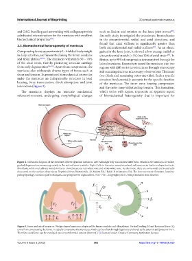
Figure 2. Schematic diagram of the structure of heterogeneous meniscus. Left: Although fully vascularized after born, vessels in the meniscus are under
gradual degeneration, remaining merely in the red-red zone in adults. Right: Cells in the outer, vascularized red-red zone are in fusiform shape similar to
fibroblasts, while oval cells are found similar to chondrocytes in red-white zone and white-white zone. Furthermore, there are some small and round cells
discovered on the surface of meniscus. Reprinted from Biomaterials, 32, Makris EA, Hadidi P, Athanasiou KA, The knee meniscus: Structure-function,
pathophysiology, current repair techniques, and prospects for regeneration, 7411–7431., Copyright (2011), with permission from Elsevier.
Figure 3. Force analysis of meniscus. Wedge-shaped meniscus adapts well to femur condyles and tibia plateau. Vertical loading (F) and horizonal force (F )
r
come from compressing the femur. F radially compresses the meniscus, which can be offset through ligaments anchored at the anterior and posterior horn.
r
Therefore, axial force can be translated into circumferential tension (from ref. [72] licensed under Creative Commons Attribution license).
Volume 9 Issue 3 (2023) 362 https://doi.org/10.18063/ijb.693