
Page 375 - IJB-9-3
P. 375
International Journal of Bioprinting 3D-printed anistropic meniscus
A C
B
D
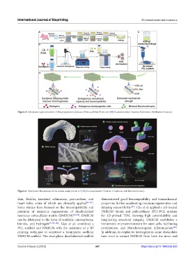
Figure 5. Schematic representation of the preparation process of the scaffolds (from ref. [88] licensed under Creative Commons Attribution license).
Figure 6. Schematic illustration of the whole study (from ref. [92] licensed under Creative Commons Attribution license).
skin, bladder, intestinal submucosa, pericardium, and demonstrated good biocompatibility and biomechanical
heart valve, some of which are clinically applied [99-101] . properties, further accelerating meniscus regeneration and
Some studies have focused on the biocompatibility and delaying osteoarthritis [107] . Cha et al. applied a cell-loaded
potential of meniscus regeneration of decellularized DMECM bioink and polyurethane (PU)-PCL mixture
meniscus extracellular matrix (DMECM) [99,102] . DMECM for 3D-printed TEM, showing high controllability and
can be fabricated in the form of scaffolds, microspheres, long-lasting structural integrity. DMECM establishes a
bioinks, and hydrogels [99,103-106] . Guo et al. combined a biomimetic microenvironment for stem cells, facilitating
PCL scaffold and DMECM with the assistance of a 3D proliferation, and fibrochondrogenic differentiation [108] .
printing technique to construct a biomimetic acellular In addition, to display its heterogeneity, some researchers
DMECM scaffold. This dual-phase decellularized scaffold have tried to extract DMECM from both the inner and
Volume 9 Issue 3 (2023) 367 https://doi.org/10.18063/ijb.693