
Page 373 - IJB-9-3
P. 373
International Journal of Bioprinting 3D-printed anistropic meniscus
A B C D
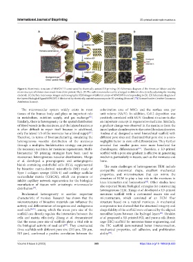
Figure 4. Biomimetic structure of MWCNT-S constructed by electrically assisted 3D printing. (A) Schematic diagram of the American lobster and the
microstructure of lobster claws made from chitin protein fibers. (B) The carbon nanotubes can be arranged in different directions by adjusting the rotating
electrode. (C) Surface microscope images and tomographic SEM images of different arrays of MWCNT-S corresponding to (B). (D) Schematic diagram of
the layered biological ligand MWCNT-S fabricat ed by electrically assisted nanocomposite 3D printing (from ref. [74] licensed under Creative Commons
Attribution license).
The microvascular system widely exists in most colonization area of MSCs and the surface area per
tissues of the human body and plays an important role unit volume (SA/V). In addition, Col-2 deposition was
in metabolism, nutrition supply, and gas exchange . positively correlated with SA/V. Gradient structure is also
[76]
Similarly, there is heterogeneity in the spatial distribution an important concept in regenerative medicine. Similarly,
of blood vessels in the meniscus, and the injured meniscus a gradient change was observed in the meniscus from the
is often difficult to repair itself because in adulthood, inner hyaline chondrocytes to the outer fibrochondrocytes.
only the lateral 1/3 of the meniscus has a blood supply . Andrea et al. designed a novel hierarchical scaffold with
[21]
Therefore, in terms of biomanufacturing, simulating the different pore sizes and illustrated that pore size is a non-
heterogeneous vascular distribution of the meniscus negligible factor in stem cell differentiation. They further
through a multiplex biofabrication strategy can provide revealed that smaller pores were more beneficial for
the necessary nutrition for meniscus regeneration. Multi- chondrogenic differentiation . Therefore, a 3D-printed
[81]
biomaterial 3D printing strategies have been used to scaffold with a pore size gradient is effective in generating
reconstruct heterogeneous vascular distributions. Margo medicine, particularly in tissues, such as the meniscus and
et al. developed a proangiogenic and antiangiogenic bone.
bioink containing endothelial cells (ECs), supplemented The main challenges of heterogeneous TEM include
by bioactive matrix-derived microfibrils (MF) made of compatible anatomical shape, excellent mechanical
Type I collagen sponge (COL-1) and cartilage acellular properties, and microstructure that can mimic the
extracellular matrix (CdECM), which can promote or structure of ECM to play a key role in the meniscus in
inhibit capillary network regeneration for the biological knee kinematics and homeostasis . Other studies have
[82]
manufacture of tissues with anisotropic microvascular also reported bionic biological strategies for constructing
distribution . heterogeneous TEM. Thiago et al. developed a 3D-printed
[77]
Biochemical heterogeneity is another important meniscus scaffold with a customized macro size and
characteristic of menisci. Research has shown that the microstructure, which consisted of an ECM fiber
microstructures of bioactive materials can influence the structure based on a natural meniscus. A mechanical
activity and differentiation of exogenous and endogenous compression test showed that the structural integrity and
seed cells [78,79] , among which the mean pore size of the shape fidelity of the scaffold were enhanced by the aligned
scaffold can directly regulate the interaction between the nanofiber layers between the hydrogel layers . Ibrahim
[83]
cells and matrix effectively. Zhang et al. demonstrated et al. proposed a 3D-printed PCL and porous silk fibroin
that the mean pore size of scaffolds plays a vital role in cage (EIC) scaffold for meniscus tissue engineering, and
the biological activity of seed cells . They constructed the EIC scaffold demonstrated better interconnection,
[80]
three scaffolds with different pore size (215 μm, 320 μm, mechanical properties, cell adhesion, and proliferation
515 μm), confirmed a positive correlation between the ability .
[84]
Volume 9 Issue 3 (2023) 365 https://doi.org/10.18063/ijb.693