Page 372 - IJB-9-3
P. 372

International Journal of Bioprinting                                       3D-printed anistropic meniscus


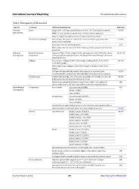
            Table 1. Heterogeneity of the meniscus
             Aspects      Contents            Relevant manifestation                                 Reference
            Anatomy       Shape               Length: 40.5 – 45.5 mm (medial meniscus), 32.4 – 35.7 mm (lateral meniscus)  [30,31]
            heterogeneity                     Width: 27 mm (medial meniscus), 26.6 – 29.3 mm (lateral meniscus)
                                              Shape: C-shaped (medial meniscus), O-shaped (lateral meniscus)
                          Vessel, nerve and lymph  Red-red zone: the outer 10 – 25% of the meniscus which is penetrated with   [13,21]
                                              vessels, nerves and lymph
                                              Red-white zone: the transitioning area                   [2,3]
                                              White-white zone: the inner 1/3 of the meniscus which is avascular, aneural and
                                              alymphatic
            Biological    Extracellular matrix   In general: Water (72%), collagens (22%), proteoglycans (1%), DNA (2%), elastin   [24,41-44]
            heterogeneity  components         (<1%) and adhesion glycoproteins (<1%). Proportion varies with age, lesions and
                                              pathological states.
                          Collagen            Red-red zone: Collagen I (80% of dry weight), Collagen II, III, IV, VI, XVIII   [50–52]
                                              (<1% of dry weight)
                                              White-white Zone: Collagen I (40% of dry weight), Collagen II (60% of dry
                                              weight)
                                              Collagens are hierarchically aligned, with randomly at superficial layer,   [23,53]
                                              circumferentially in deeper layer with radial fibers interwoven in the meniscus.
                          Cell phenotype      Fusiform fibroblast-like cells in the outer zone while oval chondrocyte-like cells   [47,48]
                                              in the inner zone with respective biological actions.
                                              Fibrochondrocytes in the peripheral region better adapt to circumferential   [49]
                                              tension while those in the inner region better withstand compressive force.
            Biomechanical   Compression       At low strain:  Circumferential: 10 MPa                  [27]
            Heterogeneity                                   Radial: 13 MPa
                                                            Axial: 19 MPa
                                              At high strain:  Circumferential: 288 MPa
                                                            Radial: 287 MPa
                                                            Axial: 299 MPa
                                              (Axial stiffness is significantly greater than both circumferential and radial stiffness)
                                              Inner zone bears compression while outer zone withstands tension.  [62,63]
                          Tension             Circumferential  Medial anterior: 99.4 MPa              [42,66]
                                                            Medial center: 107.9 MPa
                                                            Medial posterior: 114.1 MPa
                                                            Lateral anterior: 99.8 MPa
                                                            Lateral center: 78.4 MPa
                                                            Lateral posterior: 116.2 MPa
                                              Radial        Medial anterior: 10.5 MPa
                                                            Medial center: 7.6 MPa
                                                            Medial posterior: 2.5 MPa
                                                            Lateral anterior: 10.9 MPa
                                                            Lateral center: 10.3 MPa
                                                            Lateral posterior: 8.5 MPa
                                              Circumferential tensile modulus is 10-fold higher than radial modulus.
                                              Radial Young’s modulus is remarkably higher in the anterior region than   [65]
                                              posterior region.
                                              The highest tensile strength in medial meniscus appears in the posterior region.  [65]
                                                                           −14
                                                                      −15
                          Hydromechanics      Hydraulic permeability ranges from 10  to 10  m /N s for confined   [68]
                                                                              4
                                                               −15
                                                                  4
                                                          −17
                                              compression and 10  – 10  m /N s for indentation.
                                              Heterogeneity is also found circumferentially and radially between pars   [69]
                                              intermedia and posterior horn in fluid phase.
            Volume 9 Issue 3 (2023)                        364                          https://doi.org/10.18063/ijb.693
   367   368   369   370   371   372   373   374   375   376   377