
Page 448 - IJB-9-4
P. 448
International Journal of Bioprinting Single-step bioink deposition and maturation of human epidermis
2.8.2. Formation of collagen-human skin equivalent were stained with standard hematoxylin and eosin (H&E)
(c-HSE) protocol to observe cell morphology. Images were obtained
Primary keratinocytes (5.31 × 10 cells/insert) were first on a Zeiss Axio Imager Microscope (Zeiss, Germany).
6
suspended in chilled PDβ media containing NaOH and 2.10. Immunohistochemistry
then mixed with chilled bovine collagen type I, and a small
volume of this bioink was then deposited onto the indent Immunofluorescence (IF) staining was used to detect
of HDF-dermis (Figure 2). The bioink was allowed to gel at keratinocyte proliferation and differentiation. For IF
37°C for 30 min, and PDβ medium was added to the inserts. staining of primary keratinocytes, cells were briefly fixed
The next day, medium was refreshed, and the inserts were with 4% paraformaldehyde (4% PFA) for 15 min, rinsed
st
cultured submerged for another 48 h before being cultured with phosphate-buffered solution (1×) (PBS, 1 Base,
at the air-liquid interface for another 7 days before fixation Singapore), blocked with 10% donkey serum (Sigma, USA)
and staining. Media were refreshed on alternate days. and incubated at 4°C overnight with primary antibodies.
Slides were then rinsed and incubated with secondary
2.9. Hematoxylin and eosin staining antibodies and Hoechst (Sigma, USA) for 60 min, rinsed
Histological studies were performed on the HSE and c-HSE with PBS and mounted with Prolong-Gold Anti-fade
generated. Samples were fixed in 10% neutral buffered reagent (Invitrogen, USA). Confocal images were captured
formalin (NBF) for paraffin embedding. Paraffin sections on the Zeiss LSM700 (Zeiss, Germany).
A B
C D E
F
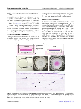
Figure 2. Dermis formation. (A) Schematic diagram showing HSE construct cultured submerged in PDβ media initially, followed by (B) culturing at airlift
interface for epidermis maturation. (C) Silicone disk used to create an indent in the collagen. (D) Transwell insert containing the transparent disk and
gelled opaque collagen. (E) Media (pink) contained within the indent on the collagen dermis. (F) H&E stained dermis showing monolayer of HDF (dark
purple) lining the indent surface and HDF (arrows) within the collagen dermis. Scale bar: 100 µm.
Volume 9 Issue 4 (2023) 440 https://doi.org/10.18063/ijb.738