
Page 446 - IJB-9-4
P. 446
International Journal of Bioprinting Single-step bioink deposition and maturation of human epidermis
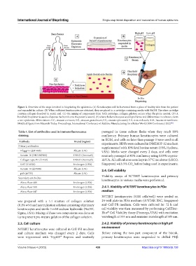
Figure 1. Overview of the steps involved in bioprinting the epidermis. (A) Keratinocytes will be harvested from a piece of healthy skin from the patient
and expanded in culture. (B) When sufficient keratinocytes are obtained, they are placed in a cartridge containing media with NaOH. The other cartridge
contains collagen dissolved in acetic acid. (C) On mixing of components from both cartridges, collagen gelation occurs when the pH is neutral. (D) A
handheld bioprinter is used to dispense the bioink onto the patient’s wound, (E) where the keratinocytes would proliferate and differentiate in culture to form
a neo-epidermis. Abbreviations: S.C., stratum corneum; S.G., stratum granulosum; S.S., stratum spinosum); S.B., stratum basale; B.M., basement membrane.
Modified figure from Materials Today: Proceedings, International Conference of Additive Manufacturing for a Better World (AM Conference) 2022 .
[42]
Table 1. List of antibodies used in immunofluorescence passaged in tissue culture flasks when they reach 80%
staining. confluency. Primary human keratinocytes were cultured
in KGM, and cells no later than passage 3 were used in all
Antibody Brand (region) experiments. HDFs were cultured in DMEM/F-12 medium
Primary antibodies supplemented with 10% fetal bovine serum (FBS, Hyclone,
Filaggrin (ab81468) Abcam (UK) USA). Media were changed every 2 days, and cells were
Keratin 10 (DKO.M7002) DAKO (Denmark) routinely passaged at 80% confluency using 0.05% trypsin-
Collagen type IV (CIV22) DAKO (Denmark) EDTA. All cell cultures were kept in 37°C incubator (ESCO,
Ki67 (514520) Invitrogen (USA) Singapore) with 5% CO before being used in experiments.
2
Keratin 14 (ab7800) Abcam (UK) 2.4. Cell viability
p63 (ab735) Abcam (UK)
Secondary antibodies Viability assays of N/TERT keratinocytes and primary
keratinocytes in various media were performed.
Alexa Fluor 488 Invitrogen (USA)
Alexa Fluor 568 Invitrogen (USA) 2.4.1. Viability of N/TERT keratinocytes in PDα
Alexa Fluor 647 Invitrogen (USA) medium
N/TERT keratinocytes (5000 cells/well) were seeded on
was prepared with a 1:1 mixture of collagen solution 24-well plate in PDα medium (A*STAR RSC, Singapore)
(0.1% w/v) and neutralization solution consisting of primary and CnT-PR medium. Cells were cultured for 72 h and
keratinocytes and sterile 5 mM sodium hydroxide (NaOH, cell viability was then measured by performing CellTiter-
®
Sigma, USA). Mixing of these two components was done on Blue Cell Viability Assay (Promega, USA) with excitation
ice to prevent pre-mature gelation of the collagen solution. wavelength at 555 nm and emission wavelength at 585 nm.
2.3. Cell culture 2.4.2. Viability of primary keratinocytes in high pH
environment
N/TERT keratinocytes were cultured in CnT-PR medium
and culture medium was changed every 2 days. Cells Before mixing the two-part component of the bioink,
were trypsinized with TrypLE™ Express and routinely primary keratinocytes were suspended in chilled PDβ
Volume 9 Issue 4 (2023) 438 https://doi.org/10.18063/ijb.738