
Page 452 - IJB-9-4
P. 452
International Journal of Bioprinting Single-step bioink deposition and maturation of human epidermis
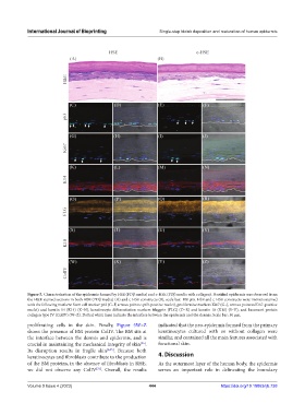
Figure 5. Characterization of the epidermis formed by HSE (PDβ media) and c-HSE (PDβ media with collagen). Stratified epidermis was observed from
the H&E stained sections in both HSE (PDβ media) (A) and c-HSE constructs (B), scale bar: 100 µm. HSE and c-HSE constructs were immunostained
with the following markers: Stem cell marker p63 (C–F, arrows point to p63-positive nuclei), proliferative markers Ki67 (G–J, arrows point to Ki67-positive
nuclei) and keratin 14 (K14) (K–N), keratinocyte differentiation markers filaggrin (FLG) (O–R) and keratin 10 (K10) (S–V), and basement protein
collagen type IV (ColIV) (W–Z). Dotted white lines indicate the interface between the epidermis and the dermis. Scale bar: 50 µm.
proliferating cells in the skin. Finally, Figure 5W–Z indicated that the neo-epidermis formed from the primary
shows the presence of BM protein ColIV. The BM sits at keratinocytes cultured with or without collagen were
the interface between the dermis and epidermis, and is similar, and contained all the main features associated with
crucial in maintaining the mechanical integrity of skin . functional skin.
[35]
Its disruption results in fragile skin [6,35] . Because both
keratinocytes and fibroblasts contribute to the production 4. Discussion
of the BM proteins, in the absence of fibroblasts in RHE, As the outermost layer of the human body, the epidermis
we did not observe any ColIV . Overall, the results serves an important role in delineating the boundary
[31]
Volume 9 Issue 4 (2023) 444 https://doi.org/10.18063/ijb.738