
Page 237 - IJB-9-5
P. 237
International Journal of Bioprinting Hydrogels for 3D bioprinting
temperature-sensitive and high-viscosity properties, but was observed, proving that local treatment is feasible.
also simulates the tissue-specific microenvironments However, this study failed to point out the mechanism by
of cartilage and vascularized fibrous tissue. Because the which ECM promotes local treatment response. But we
alternating soft and hard tissue structure of the stent is can still get some enlightenment. By extracting the tissue-
very close to the human body’s own trachea, functional specific EdECM hydrogel from the esophageal ECM, it can
reconstruction of the mechanical and physiological produce local treatment without the use of drugs. That is,
properties of the trachea has been successfully achieved. the dECM hydrogel obtained on a specific site is essential
Hong et al. used glycidyl methacrylate (GMA) to for tissue repair.
[34]
modify the chondrocyte-loaded SF hydrogel (Silk-
GMA) for DLP-based printing. Research and testing In urethral repair tissue engineering, its structure is
showed that the scaffold has a powerful mechanical similar to that of the trachea. The construction methods
advantage effect on the regeneration of defective tissues. of 3D bioprinting technology can be used as reference
At the same time, in vivo experiments were carried out methods. Zhang et al. [146] designed a “sandwich” tubular
on a rabbit model with a partially defective trachea, and structure. Using PCL and poly (lactide-caprolactone-
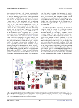
new cartilage tissue and epithelium (Figure 7B) were cocaprolactone) (PLCL) thermoplastic polymer and cell-
found around the transplanted Silk-GMA hydrogel. The loaded fibrin, gelatin, and other hydrogels realizes the
esophagus is next to the trachea, which also has strictures 3D-bioprinted bionic urethral structure for the first time.
and inflammation. Currently, there is no direct treatment They first obtained the structural index of the rabbit urethra
method for patients with radiation esophagitis. Ha et and then used a spiral stent made of a blend of PCL and
al. [148] proposed a pioneering direct treatment strategy. PLCL (50:50 ratio) as the intermediate layer. Three layers
They developed an esophageal-derived dECM (EdECM) of the smooth muscle cells (SMCs)-loaded hydrogel are
hydrogel and then used a rotating rod combined with printed on the outer layer of the scaffold, and two layers of
3D printing system to prepare an EdECM hydrogel stent. the urothelial cells (UCs)-loaded hydrogel are printed on
They verified that EdECM hydrogel has good rheological the inner layer. The results showed that the 3D-bioprinted
properties and biological functions. Besides, in a rat model PCL/PLCL scaffold has mechanical properties comparable
of radiation esophagitis, the therapeutic effect of the stent to those of the natural rabbit urethra. The fibrin hydrogel
Figure 7. (A) Schematic diagram of in situ printing; experimental comparison of in situ printing on mouse skin. Reproduced with permission [158] . (B) DLP-
printed silk-GMA hydrogel to prepare tracheal stents, which were used in rabbit models to promote the regeneration of cartilage tissue and epithelium.
Reproduced with permission [31]. (C) The first ever printed organ was a heart printed in a suspended medium. Reproduced with permission [159] . (D) Flow
chart of 3D printing kidney phantom. Reproduced with permission [160] .
Volume 9 Issue 5 (2023) 229 https://doi.org/10.18063/ijb.759