
Page 251 - IJB-9-5
P. 251
International Journal of Bioprinting
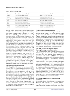
Table 1. Primers used in RT-PCR
Gene name Forward primer sequence (5’ to 3’) Reverse primer sequence (5’ to 3’)
NCAD CACCCGGCTTAAGGGTGATT CGATCCTGTCTACGTCGGTG
NCAN GTGAAGTCAGCCATCCACCA TCGGGACGAGTGTAGAGTGT
Integrin β1 ACTCAGTGAACAGCAACGGTGAAG TCCAAATCAGCAGCAAGGCAAGG
Cdc42 GCTGTCAAGTATGTGGAGTGT GGCTCTGGAGATGCGTTCA
Rac1 CAATGCGTTCCCTGGAGAGT AACACGTCTGTTTGCGGGTA
MPZ CTGCCCTGCTCTTCTCTTCTTTGG GAGCCCACAGCACCATAGACTTC
PMP22 GGTGCTAGTGTTGCTCTTCGTCTC CAAGGCGGATGTGGTACAGTTCTG
GAPDH AACTTTGGCATCGTGGAAGG TGGATGCAGGGATGATGTTCTG
printing system. We set the experimental parameters 2.10. Immunofluorescence staining
as follows: the exposure intensity was 80 mW/cm , Cells were inoculated on the samples and cultured as
2
the exposure time was 5 s, and the thickness was 0.1 mm. described above. After 48 h, the samples were fixed in 4%
We selected ASF-MA 10% with the highest degree of paraformaldehyde in PBS and permeabilized with 0.5%
methacryloylation; the appropriate concentration was Triton X-100 for 20 min. The samples were then blocked with
20 wt%; the LAP concentration was 1 wt%. To print the normal goat serum for 30 min for non-specific binding. The
“peace dove,” we mixed the ASF-MA bioinks with the rabbit adherent spot protein antibody (1:100) was incubated
red fluorescent protein-labeled mouse colon cancer cells overnight at 4°C. The next day, Alexa Fluor® 488-labeled
(CT26-RFP). We placed the bioinks with cells on a circular goat anti-rabbit IgG (1:50), rhodamine ghost pen cyclic
slide with a diameter of 16 mm for bioprinting. Similarly, to peptide (1:50), and DAPI PBS solution were added and
print the “Yin-Yang Tai Chi” diagram, we mixed the ASF- incubated at room temperature, and all mounted samples
MA bioinks with CT26-RFP cells and GFP-labeled mouse were visualized by confocal laser scanning microscopy.
bone marrow stromal cells (OP9-GFP), respectively, and
printed it twice. We placed the bioinks with cells on a 2.11. RNA isolation and real-time reverse
circular slide with a diameter of 16 mm for bioprinting. transcription-polymerase chain reaction
We used sterile PBS to wash to remove uncrosslinked The mRNA expression levels of related genes including
monomers and other impurities. Then, we continued to NACD, Integrin β1, Cdc42, Rac1, MPZ, and PMP22
print on the slide for the second time. After printing, we were examined using real-time reverse transcription-
transferred the sample to the 12-well plate. We used sterile polymerase chain reaction (RT-PCR), and glycerol
PBS to wash 6 times for 5 min each time. After 15 min, triphosphate dehydrogenase (GAPDH) was selected as the
we replaced PBS with Dulbecco’s Modified Eagle Medium. housekeeping gene. RT-PCR procedure was performed
We placed the 12-well plate in incubator. Finally, on the 1, as follow: First, cellular RNA was extracted using a total
2, and 3 days, we observed the cell state and recorded the RNA extraction kit. The RNA concentration of each group
fluorescence intensity under the fluorescence microscope. was measured using a nucleic acid and protein analyzer,
and the RNA was stored in a -80°C freezer. Next, reverse
2.9. Cytocompatibility of hydrogel transcription was performed using the extracted RNA
ASF-MA 10% (20 wt%) was used for the printing bioinks. as template RNA. The reverse transcription reaction was
S16 cells, NIH/3T3 cells, and HUVECs in the logarithmic performed in a PCR machine. The cDNA was obtained
growth phase were inoculated at a density of 8 × 10 after the reaction and stored in a -20°C refrigerator for
2
cells/well, 1 × 10 cells/well, and 1 × 10 cells/well in the subsequent experiments. Finally, using GAPDH as an
3
3
96-well plate, respectively. After 1, 3, 5, and 7 days of internal reference, the mRNA expression levels of NCAD,
culture, the cell growth and proliferation on the hydrogels Integrin β1, Cdc42, Rac1, MPZ, and PMP22 were analyzed
were assessed by using CCK-8 assay according to the by RT-PCR. All primers used are listed in Table 1.
protocol. The S16 cells, NIH/3T3 cells, and HUVECs in
the logarithmic growth phase were respectively seeded 2.12. In vivo degradation test and histological
onto the printed hydrogel in a 12-well plate at a density of evaluation
2 × 10 cells/well and cultured in an incubator. After 1, 3, Cylindrical hydrogels (6 mm diameter, 2 mm height) were
4
and 5 days of incubation, live/dead assays were employed fabricated from sterile ASF-MA 10% , GelMA, and BSF-
to examine the viability, and the cells were photographed GMA printing inks by 3D printing. We used SPF-grade
under a fluorescence microscope. BALB/c male mice for dorsal subcutaneous implantation
Volume 9 Issue 5 (2023) 243 https://doi.org/10.18063/ijb.760