
Page 252 - IJB-9-5
P. 252
International Journal of Bioprinting Antheraea pernyi silk fibroin bioinks for DLP 3D printing
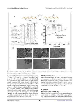
Figure 1. (A) The synthetic route of ASF-MA. (B) The H-NMR spectra of ASF and ASF-MA. (C) Standard curve and the content of free amino groups of
1
ASF-MA. (D) The SEM images of the different ASF-MA hydrogels.
experiments. First, mice were anesthetized using approved 2.13. Statistical analysis
procedure. The back of the mouse was shaved and Three independent experiments with three parallel samples
disinfected. Three wounds of 2 cm were cut on the back per experiment were undertaken, unless otherwise stated.
skin with sterilized surgical instruments, and the skin was The data are expressed as mean ± standard deviation (SD).
partially separated. ASF-MA 10% , GelMA, and BSF-GMA The statistical difference was analyzed by one-way analysis
hydrogels were implanted subcutaneously on the back. The of variance (ANOVA) using the GraphPad Prism software
back skin of the mouse was sutured with 8-0 sutures. The (ns: p > 0.05, *p < 0.05, **p < 0.01, ***p < 0.001).
mice were euthanized at pre-determined time points (1, 2,
3, 4, 8, 10, and 12 weeks) after transplantation to harvest 3. Results
the transplanted hydrogels together with the surrounding
tissues. We weighed the transplanted hydrogel after 3.1. Characteristics of ASF-MA
removing the outer fibrous capsule and surface liquid for The ASF-MA bioink was prepared by reacting MA with free
record. The degradation rate was shown as the ratio of wet amino groups on ASF (Figure 1A). While preparing ASF-
weight to initial wet weight at each time point. MA, ASF would coagulate and could not be reconstituted
Volume 9 Issue 5 (2023) 244 https://doi.org/10.18063/ijb.760