
Page 256 - IJB-9-5
P. 256
International Journal of Bioprinting Antheraea pernyi silk fibroin bioinks for DLP 3D printing
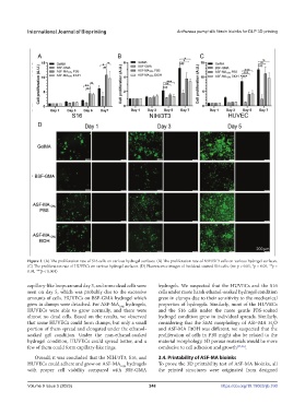
Figure 3. (A) The proliferation rate of S16 cells on various hydrogel surfaces. (B) The proliferation rate of NIH/3T3 cells on various hydrogel surfaces.
(C) The proliferation rate of HUVECs on various hydrogel surfaces. (D) Fluorescence images of live/dead stained S16 cells. (ns: p > 0.05, *p < 0.05, **p <
0.01, ***p < 0.001)
capillary-like loops around day 3, and some dead cells were hydrogels. We suspected that the HUVECs and the S16
seen on day 5, which was probably due to the excessive cells under more harsh ethanol-soaked hydrogel condition
amounts of cells. HUVECs on BSF-GMA hydrogel which grew in clumps due to their sensitivity to the mechanical
grew in clumps were detached. For ASF-MA 10% hydrogels, properties of hydrogels. Similarly, most of the HUVECs
HUVECs were able to grow normally, and there were and the S16 cells under the more gentle PBS-soaked
almost no dead cells. Based on the results, we observed hydrogel condition grew in individual spreads. Similarly,
that some HUVECs could form clumps, but only a small considering that the SEM morphology of ASF-MA H O
2
portion of them spread and elongated under the ethanol- and ASF-MA EtOH was different, we suspected that the
soaked gel condition. Under the non-ethanol-soaked proliferation of cells in PBS might also be related to the
hydrogel condition, HUVECs could spread better, and a material morphology. 3D porous materials would be more
few of them could form capillary-like rings. conducive to cell adhesion and growth [35,36] .
Overall, it was concluded that the NIH/3T3, S16, and 3.4. Printability of ASF-MA bioinks
HUVECs could adhere and grow on ASF-MA 10% hydrogels To prove the 3D printability test of ASF-MA bioinks, all
with proper cell viability compared with BSF-GMA the printed structures were originated from designed
Volume 9 Issue 5 (2023) 248 https://doi.org/10.18063/ijb.760