
Page 260 - IJB-9-5
P. 260
International Journal of Bioprinting Antheraea pernyi silk fibroin bioinks for DLP 3D printing
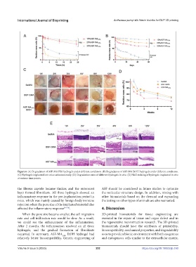
Figure 6. (A) Degradation of ASF-MA PBS hydrogels under different conditions. (B) Degradation of ASF-MA EtOH hydrogels under different conditions.
(C) Hydrogels implanted into mice subcutaneously. (D) Degradation rates of different hydrogels in vivo. (E) H&E staining of hydrogels implanted in vivo
at various time points.
the fibrous capsule became thicker, and the outermost ASF should be considered in future studies to optimize
layer formed fibroblasts. All three hydrogels showed an the molecular structure design. In addition, mixing with
inflammatory response in the pre-implantation period in other biomaterials based on the demand and expanding
mice, which was mainly caused by foreign body immune the testing on other types of animals are also warranted.
rejection when the pore size of the implanted material also
affected the inflammatory response [44-46] . 4. Discussion
When the pore size became smaller, the cell migration 3D-printed biomaterials for tissue engineering are
rate and cell infiltration rate would be slow. As a result, essential in the repair of tissue and organ defect and in
we could see the enhancement of the inflammation. the regenerative reconstruction research. The 3D-printed
After 2 months, the inflammation resolved on all three biomaterials should have the attributes of printability,
hydrogels, and the gradual formation of fibroblasts biocompatibility, mechanical properties, and degradability
occurred. In summary, ASF-MA 10% EtOH hydrogel had so as to provide a bionic environment with both exogenous
relatively better biocompatibility. Genetic engineering of and endogenous cells similar to the extracellular matrix.
Volume 9 Issue 5 (2023) 252 https://doi.org/10.18063/ijb.760