
Page 272 - IJB-9-5
P. 272
International Journal of Bioprinting 3D-printed scaffolds for TMJ fibrocartilage regeneration
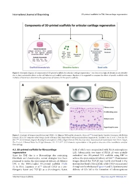
Figure 2. Schematic diagram of components of 3D-printed scaffolds for articular cartilage regeneration. *, too low or too high cell density is not advisable
due to their undesirable effects on the cell behavior and scaffold performance; therefore, it is suggested to compare the effects of specific scaffolds with
multiple cell densities to determine the appropriate cell density for the specific scaffolds.
Figure 3. Anatomy of temporomandibular joint (TMJ). (A) Human TMJ sagittal schematic. (from ref. licensed under Creative Commons Attribution
[95]
license). (B) A 3D computer-aided design model of human TMJ. Reproduced with permission from Legemate K, Tarafder S, Jun Y, et al., J Dent Res, 95:
800–7 . (C) Schematic diagram showing the arrangement of the collagen fibers in TMJ disc (top view). Reproduced with permission from She Y, Tang S,
[75]
Zhu Z, et al., J Biomed Mater Res B Appl Biomater, 111: 717–29 . (D) Schematic representation of the gradient structure of the mandibular condylar
[96]
fibrocartilage.
4.2. 3D-printed scaffolds for fibrocartilage both of which were encapsulated with PLGA microspheres
regeneration (μS). Subsequently, two types of PLGA μS were spatially
Since the TMJ disc is a fibrocartilage disc containing embedded into 3D-printed PCL scaffolds using EBP to
fibroblasts and chondrocytes, several strategies have been achieve the spatiotemporal delivery of GFs . Fluorescence
[76]
proposed to realize the spatiotemporal delivery of different images showed that TGF-β3 was mainly distributed in the
GFs in the MSCs-loaded 3D-printed scaffolds (Table intermediate band of the scaffold, while CTGF was scattered
2). In 2016, Legemate et al. selected CTGF as a pro- throughout the whole area (Figure 4A and B). Spatiotemporal
[75]
fibrogenic factor and TGF-β3 as a chondrogenic factor, delivery of GFs led to the formation of inhomogeneous
Volume 9 Issue 5 (2023) 264 https://doi.org/10.18063/ijb.761