
Page 275 - IJB-9-5
P. 275
International Journal of Bioprinting
coated the PCL/PU scaffolds with polydopamine (PDA)
and then combined them with dECM derived from porcine
TMJ discs (Figure 4E). The modified scaffolds exhibited Bioactive factors
higher tensile modulus and compressive moduli. PDA-PCL/
PU and PDA-PU had similar compressive modulus to the BMP-7 BMP-2
central region and the peripheral region of the human TMJ - - - -
disc, respectively. Compared with the original scaffolds,
the chondrogenic-specific markers (Sox 9 and Col II) and
fibrous-specific marker (Col I) were upregulated in the Scaffold materials
modified scaffolds after they were seeded with rat costal
chondrocytes and L929 fibroblasts and cultured for 14 days. PLA PCL* PCL/HA PGA/PLA PCL Gelatin**
In vivo tests further confirmed the ability of the PDA coating HA HA
to enhance chondrogenesis and fibrogenesis.
5. Mandibular condyle cartilage tissue 2.5 × 10 7 cells/mL 2 × 10 5 cells/mL; 1.2 × 10 6 cells/mL
engineering Cell density 5 × 10 7 cells/mL 5 × 10 7 cells/mL
5.1. Anatomy - - -
The immunohistochemical staining of the fibrocartilage
of the mandibular condyle revealed that type I and type
II collagen predominate in the superficial zone and deep
zone (the mature and hypertrophic zones), respectively, Pig chondrocytes Minipig chondrocytes
[81]
which is different from articular hyaline cartilage . Minipig BMSCs Rat BMSCs
Specifically, the fibrocartilage covering the upper surface Cell type HGFs
of the mandibular condyle can be subdivided into four - - -
layers super-inferiorly: fibrous, proliferating, mature,
and hypertrophic zones, where the fiber organization Abbreviations: SLS, selective laser sintering; PCL, polycaprolactone; HA, hydroxyapatite; HGFs, human gingival fibroblasts; FDM, fused deposition modeling; CS, chitosan; PGA/PLA, polyglycolic condylar head of the scaffold was packed with iliac crest bone marrow from the minipig; **, the gelatin scaffolds were crosslinked with dehydrothermal, ribose glycation, dehydrotherma
[82]
and cellular composition vary (Figure 3D) . Flat-shape Animal model acid/polylactic acid; BMSCs, bone mesenchymal stem cells; TGF-β1, transforming growth factor beta 1; BMP-2, bone morphogenetic protein 2; PLGA, poly(D, L-lactic-co-glycolic acid); *, the
fibroblasts and type I collagen occupied the fibrous zone. Minipig Minipig
MSCs, which serve as chondrocyte precursors, were Mice Sheep Mice -
distributed in the proliferative zone. The mature and
hypertrophic zones are composed of type II collagen with
loose organization and mature chondrocytes. Aggrecan
was mainly found in the mature and hypertrophic zones In vitro and in vivo
and not in the fibrous zone. The collagen fiber network Study design
and proteoglycans provide load-bearing functions to the In vivo In vivo In vivo In vivo In vitro
mandibular condyle. Singh et al. divided the mandibular
[83]
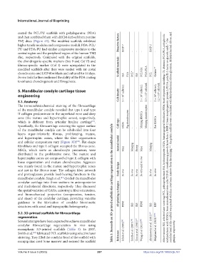
condylar cartilage into three sections in anteroposterior Table 3. Research on 3D-printed scaffolds for cartilage regeneration in the mandibular condyle
and mediolateral directions, respectively. They discussed
the spatial variation of GAGs, anisotropic fiber orientation,
and biomechanical properties (compression, tension, 3D printing techniques
and shear) of the condylar cartilage, providing valuable
guidance to the fabrication of condylar biomimetic
structures with zonal and topographic heterogeneity. FDM SLS SLS FDM SLS EBP
5.2. 3D-printed scaffolds for fibrocartilage
regeneration
Several attempts have been reported to achieve mandibular
condylar fibrocartilage regeneration in vivo using Abramowicz et al. (2021) [85]
monophasic 3D-printed scaffolds (Table 3). In 2007, Schek et al. (2005) [88] Smith et al. (2007) [84] Ciocca et al. (2013) [87] Wang et al. (2017) [89] Helgeland et al. (2021) [90,91]
[84]
Smith et al. fabricated PCL scaffolds using selective laser genipin, respectively.
sintering. They filled the condylar head of the scaffold with Reference
minipig iliac crest bone marrow and secured the scaffold
Volume 9 Issue 5 (2023) 267 https://doi.org/10.18063/ijb.761