
Page 276 - IJB-9-5
P. 276
International Journal of Bioprinting 3D-printed scaffolds for TMJ fibrocartilage regeneration
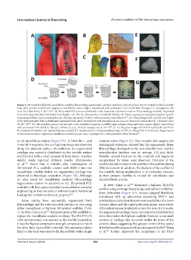
Figure 5. 3D-printed scaffolds for mandibular condylar fibrocartilage regeneration. (A) Iliac crest bone marrow packed into 3D-printed scaffold condylar
head (left) and the scaffold well adapted to mandibular ramus (right). Reproduced with permission from Smith MH, Flanagan CL, Kemppainen JM,
et al., Int J Med Robot, 3: 207–16 . (B) 3D-printed PCL porous scaffold with collar fixation fit (outlined in red) on 3D-printed pig mandible. Reprinted
[84]
from Oral Surg Oral Med Oral Pathol Oral Radiol, 132: 145–52, Abramowicz S, Crotts SJ, Hollister SJ, Tissue-engineered vascularized patient-specific
temporomandibular joint reconstruction in a Yucatan pig model, © (2021), with permission from Elsevier . (C) Visual design (left) and real view (right)
[85]
of the hydroxyapatite (HA) scaffold and customized bone plates. Reproduced with permission from Ciocca L, Donati D, Fantini M et al., J Biomater Appl,
28: 207–18 . (D) The assembly process and real view of the assembled composite scaffold (upper polymer phase and lower ceramic phase). Reproduced
[87]
with permission from Schek R, Taboas J, Hollister S, et al., Orthod Craniofac Res, 8: 313–9 . (E–G) The gross images of PCL/HA scaffold (E) and PGA/
[88]
PLA scaffold (F) and the well-matched biphasic scaffold (G). Reprinted from J Craniomaxillofac Surg, 45: 855–61, Wang F, Hu Y, He D, et al., Regeneration
of subcutaneous tissue-engineered mandibular condyle in nude mice, Copyright 2017, with permission from Elsevier .
[89]
to the mandibular ramus (Figure 5A). At both the 1- and titanium screw (Figure 5C). Four months after surgery, the
3-month time points, the cartilaginous tissue was observed histological evaluation showed that the regenerated dense
along the articular surface. Nevertheless, the regenerated fibrocartilage developed on the new articular bone and the
cartilage was unevenly distributed on the condyle surface osteochondral interface was on average 1.25-mm thick.
and blended with a small amount of bony tissue. Another Notably, several fractures in the material and fragments
similar study reported different results. Abramowicz encapsulated by tissue were observed. Fractures of the
[85]
et al. found that 6 months after implantation of scaffold are detrimental to the stability of the scaffold during
3D-printed PCL scaffolds coated with BMP-2 into the TMJ movement. In addition, the fixation of the scaffold to
mandibular condyle defect, no regenerated cartilage was the condyle during implantation is of particular concern,
observed in histologic evaluation (Figure 5B). Although as firm primary stability is crucial for osteoblastic and
an ideal result for mandibular condylar fibrocartilage chondroblastic activity.
regeneration cannot be achieved so far, 3D-printed PCL In 2005, Schek et al. fabricated a biphasic PLA/HA
[88]
scaffolds still have great potential in mandibular condylar scaffold using an image-based design and indirect solid free-
engineering as they are able to withstand early functional form fabrication (Figure 5D). Human gingival fibroblasts
[86]
loading due to their mechanical property .
transduced with an adenovirus expressing BMP-7 and
Some studies have successfully regenerated both porcine knee joints chondrocytes were seeded into the lower
fibrocartilage and the osteochondral interface in vivo using ceramic phase and the upper polymeric phase, respectively.
either monophasic or biphasic 3D-printed scaffolds (Table After subcutaneous implantation into the mice for 4 weeks,
3). In 2013, Ciocca et al. fabricated porous HA scaffolds to the regenerated cartilage, bone, and osteochondral interface
[87]
replace the mandibular condyles in sheep. The HA-F70 (70 were observed in the biphasic scaffold. However, some small
vol% total porosity) was selected as the scaffold material as pockets of cartilage also occurred within the pores of the
it had the highest compressive strength compared to that of ceramic phase, suggesting that greater control of the spatial
the other three types of HA materials. The customized plates distribution of the regenerated tissue is required. In 2017, Wang
fixed on the bone were used to fix the scaffolds with a single et al. further improved the component of the PLA/
[89]
Volume 9 Issue 5 (2023) 268 https://doi.org/10.18063/ijb.761