
Page 279 - IJB-9-5
P. 279
International Journal of Bioprinting
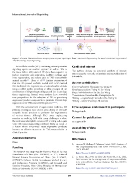
Figure 7. Schematic illustration of some emerging tissue-engineering strategies (lower row) potentially promoting the research translation (upper row) of
TMJ fibrocartilage tissue engineering.
Extracellular vesicles (EVs) containing various paracrine Conflict of interest
signaling agents are another approach to deliver BFs to
the tissue defect. MSC-derived EVs have been reported to The authors declare no potential conflicts of interest
induce progenitor cells migration, facilitate cartilage and concerning the research, authorship, and/or publication of
bone regeneration, and relieve pain in TMJ osteoarthritis this article.
[63]
animal models . Chen et al. [104] further demonstrated
that the 3D-printed scaffolds loaded with MSC-derived Author contributions
EVs facilitated the regeneration of osteochondral defects Conceptualization: Shoushan Hu, Yating Yi
using a rabbit model, providing an ideal example of the Funding acquisition: Yating Yi, Jun Wang
combination of 3D printing techniques and EVs in cartilage Project administration: Jin Liu, Jun Wang
tissue engineering. Several recent reviews have provided Visualization: Shoushan Hu, Chengxinyue Ye
new perspectives for the adoption of EVs as promising Writing – original draft: Shoushan Hu, Yating Yi
engineered product components to promote fibrocartilage Writing – review & editing: All authors
regeneration for TMJ osteoarthritis patients [105-107] .
With the advancement of regenerative medicine, 3D Ethics approval and consent to participate
printing techniques have shown great ability to fabricate Not applicable.
complex bionic products to promote the regeneration
of various tissues. Although TMJ tissue engineering
remains an evolving field with many challenges to date, Consent for publication
the continued attempts to combine 3D printing techniques Not applicable.
with TMJ tissue engineering will likely bring us closer to
a future where 3D-printed tissue-engineered products Availability of data
become an effective treatment for TMJ osteoarthritis in
clinical practice. Not applicable.
Acknowledgments References
None.
1. Alomar X, Medrano J, Cabratosa J, et al., 2007, Anatomy of
Funding the temporomandibular joint. Semin Ultrasound CT MR,
28(3): 170–183.
This research was supported by National Natural Science http://doi.org/10.1053/j.sult.2007.02.002
Foundation of China (No. 82101059) to Y.Y.; National
Natural Science Foundation of China (No. 81970967, 2. de Souza RF, Lovato da Silva CH, Nasser M, et al., 2012,
82271019), Sichuan Health Commission Medical Science Interventions for the management of temporomandibular
and Technology Program (21ZD003), and Research and joint osteoarthritis. Cochrane Database Syst Rev, 2012(4):
Develop Program, West China Hospital of Stomatology Cd007261.
Sichuan University (RD-03-202101) to J.W. http://doi.org/10.1002/14651858.CD007261.pub2
Volume 9 Issue 5 (2023) 271 https://doi.org/10.18063/ijb.761