
Page 274 - IJB-9-5
P. 274
International Journal of Bioprinting 3D-printed scaffolds for TMJ fibrocartilage regeneration
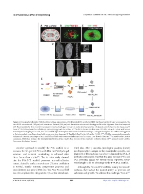
Figure 4. 3D-printed scaffolds for TMJ disc fibrocartilage regeneration. (A) 3D-printed PCL scaffolds of TMJ disc based on the 3D laser scanning data. The
size of PCL microstrands (300 μm) and interstrand distance (300 μm) and the relative microstrand density parallel to the alignment direction compared
with the perpendicular direction (2:1) were determined to closely approximate the native tensile properties. (B) Spatial control of connective tissue growth
factor (CTGF) throughout the scaffolds and transforming growth factor beta 3 (TGF-β3) in the intermediate zone. (C) After a 6-week culture with human
mesenchymal stem/progenitor cells, the CTGF and TGF-β3 microsphere-embedded scaffold showed type I collagen throughout the scaffold and aggrecan
[75]
in the intermediate zone. Reproduced with permission from Legemate K, Tarafder S, Jun Y, et al., J Dent Res, 95: 800–7 . (D) Microcomputed tomography
[77]
analysis and cross-section images of PCL scaffold modified with a PEGDA shell (upper) and a PEGDA core (lower). (from ref. licensed under Creative
Commons Attribution license.) (E) Schematic illustration of the construction process of the composite scaffolds. (from ref. licensed under Creative
[80]
Commons Attribution license).
Another approach to modify the PCL scaffold is to third disc. After 3 months, histological analysis showed
immerse the 3D-printed PCL scaffold in the PVA hydrogel no degenerative changes in the mandibular condyle, and
solution, and network crosslinking is achieved after ingrowth of fibrous tissue was observed around the PCL. A
three freeze-thaw cycles . The in vitro study showed probable explanation was that the gaps between PVA and
[79]
that the PVA-PCL scaffold possessed non-cell-adhesive PCL provided spaces for fibrous tissue ingrowth, which
nature, desirable surface smoothness (friction coefficient was thought to be an advantage of the PVA-PCL scaffold.
at 0.0662), similar porosity, compressive property, and Although the PCL and PU scaffolds usually had smooth
viscoelasticity as a natural TMJ disc. The PVA-PCL scaffold surfaces, they lacked the desired ability to promote cell
was then implanted in the goats to replace the lateral one- adhesion and growth. To address this challenge, Yi et al.
[80]
Volume 9 Issue 5 (2023) 266 https://doi.org/10.18063/ijb.761