
Page 273 - IJB-9-5
P. 273
International Journal of Bioprinting
fibrocartilaginous tissue in the scaffold after 6 weeks of
Bioactive factors CTGF, TGF-β3 CTGF, TGF-β3 and GAG contents were detected in the anterior/posterior
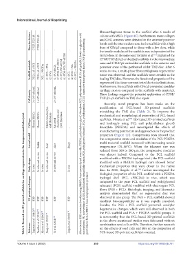
Abbreviations: EBP, extrusion-based printing; BMSCs, bone mesenchymal stem cells; PEGDA, poly(ethylene glycol) diacrylate; PCL, polycaprolactone; PLGA μS, poly(D, L-lactic-co-glycolic acid)
culture with MSCs (Figure 4C). Furthermore, more collagen
bands and the intermediate zone in the scaffolds with a high
dose of GFs/μS compared to those with a low dose, while
the tensile modulus of the scaffolds was independent of the
-
-
-
-
GF/μS dose. In the same year, Tarafder et al. implanted the
[71]
CTGF/TGF-β3/μS-embedded scaffolds in the intermediate
Scaffold materials PLGA μS + PCL PLGA μS + PCL PCL + PEGDA (1) PCL + PVA (2) PVA (1) PU-dECM (2) PDA-PU-dECM (3) PCL/PU-dECM (4) PDA-PCL/PU-dECM (1) PCL (2) PGS + PCL (3) PCL + PEGDA zone and CTGF/μS-embedded scaffolds in the anterior and
posterior areas of the perforated rabbit TMJ disc. After 6
weeks in vivo, a multi-phase fibrocartilaginous regenerative
tissue was observed, and the scaffolds were invisible in the
healing TMJ disc. However, the functional properties of the
regenerated disc tissue were not tested due to size limitations.
Furthermore, the scaffolds with GFs/μS prevented condylar
cartilage erosion compared to the scaffolds with empty/μS.
Cell density 2 × 10 6 cells/mL 1 ×10 6 cells/mL 2 × 10 6 cells/mL These findings suggest the potential application of CTGF/
TGF-β3-μS scaffolds in TMJ disc repair.
Recently, novel progress has been made on the
-
-
-
modification of PCL-based 3D-printed scaffolds
mimicking the TMJ disc (Table 2). To improve the
Table 2. Research on 3D-printed scaffolds for cartilage regeneration in the temporomandibular joint
scaffolds, Moura et al. fabricated 3D-printed scaffolds
[77]
Cell type Human BMSCs Human BMSCs Rabbit chondrocytes and fibroblasts Rat costal chondrocytes and L929 fibroblasts mechanical and morphological properties of PCL-based
and hydrogels using PCL and poly(ethylene glycol)
diacrylate (PEGDA) and investigated the effects of
-
-
manufacturing parameters and approaches on the product
properties (Figure 4D). Compression tests showed that
Animal model Rabbit microspheres; PU, polyurethane; dECM, decellularized extracellular matrix; PGS, poly(glycerol sebacate); PDA, polydopamine; CTGF, connective tissue growth factor; TGF-β3, transforming the compression stress and modulus of the PCL-PEGDA
multi-material scaffold increased with increasing nozzle
Sheep
Goat
Mice
temperature (78–86°C). When the filament size was
-
-
reduced from 300 to 200 μm, the compressive modulus
was almost halved. Compared to the PCL scaffold
Study design In vitro In vitro and in vivo In vitro In vitro and in vivo In vitro and in vivo In vivo modified with a PEGDA hydrogel shell, the PCL scaffold
modified with a PEGDA hydrogel core showed better
mechanical properties that were closer to the native
disc. In 2021, Ângelo et al. further investigated the
[78]
biological properties of the PCL scaffold with a PEGDA
growth factor beta 3; BMP-2, bone morphogenetic protein 2.
hydrogel shell (PCL +PEGDA) in vivo, which was
3D printing techniques compared to the pure PCL scaffold and poly(glycerol
sebacate) (PGS) scaffold modified with electrospun PCL
fibers (PGS + PCL). Histologic, imaging, and kinematic
analysis demonstrated that no regenerated disc was
observed in any group. The PGS + PCL scaffold showed
EBP
EBP
EBP
EBP
EBP
EBP
excellent biocompatibility as it was rapidly resorbed.
Besides, the PGS + PCL scaffold prevented condylar
degenerative changes, which were still observed in both
Legemate et al. (2016) [75] Tarafder et al. (2016) [71] Moura et al. (2020) [77] Jiang et al. (2021) [79] Ângelo et al. (2021) [78] is noteworthy that the PCL-based 3D-printed scaffolds
the PCL scaffold and PLA + PEGDA scaffold groups. It
in the above-mentioned studies were fabricated without
combination seed cells or BFs. Therefore, further research
Reference Yi et al. (2021) [80] on the effects of seed cells and BFs on the properties of
PCL-based 3D-printed scaffolds is needed.
Volume 9 Issue 5 (2023) 265 https://doi.org/10.18063/ijb.761