
Page 306 - IJB-9-5
P. 306
International Journal of Bioprinting Scaffold for engineering enthesis organ
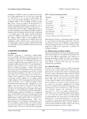
multiphasic scaffold for enthesis healing has been faced Table 1. Slicing and printing parameters.
in two main approaches: (i) the direct use of stem cells Parameters Settings Unit
(i.e., BM-MSCs), or (ii) the use of differentiated cells
such as tenocytes, tendon fibroblasts, chondrocytes, and Infill 50 %
osteoblasts (OBs) . The accessibility of MSCs prompts Layer thickness 0.2 mm
[20]
their choice; however, it requires the development of a Printing speed 12 mm/s
scaffold with regional biochemical or mechanical cues to Printing pattern* Rectilinear –
induce specific lineage differentiation in distinct zones [12,17] . Extrusion width 0.4 mm
In this context, this work aims to design and fabricate an Nozzle diameter 0.4 mm
innovative multimaterial and multiscale scaffold capable of
inducing cells into a graded enthesis-like tissue comprising Nozzle temperature 160 °C
a T/L region and a bone region. Specific fabrication Bed temperature 35 °C
methods for different biomaterials were used to obtain *Each layer is 90 degrees oriented with the previous one.
the enthesis scaffold. MSCs or pre-committed MSCs
into tenocytes and osteoblast were combined within the dissolving the polymers in chloroform (Sigma-Aldrich,
multiphasic scaffold. With the aim to push forward the St. Louis, Missouri, United States), while PCL (23% w/v)
T/L engineering, a clinically relevant construct fabricated was dissolved in acetic acid. Scaffolds were fabricated by
by braiding three enthesis scaffolds was also presented and casting the prepared solutions into Petri dishes at room
characterized. temperature until solvent evaporation to perform the
biological validation.
2. Materials and methods 2.2. Biofabrication of enthesis scaffold
2.1. Materials This section is divided into three sub-paragraphs to explain
Extensive screening of commercial, medical-grade, better the optimization of the fabrication parameters and
and bioresorbable natural and synthetic polymers was how the two different technologies were combined to
conducted to select the most valuable polymers for T/Ls fabricate the enthesis scaffold. As a result of the polymer
and enthesis engineering. The following materials were screening (see results section), the PCL and the PLGA
[21]
evaluated: (i) gelatin from porcine skin (Type A) and were selected to replicate the bone and T/Ls tissues
[22]
(ii) gelatin methacryloyl (GelMA) as natural polymers, characteristics, respectively.
[23]
(iii) poly(l-lactic acid) (PLLA) , (iv) PCL, and (v) 75:25 2.2.1. Bone-like region
PLGA as synthetic polymers . Gelatin (gel strength The PCL was processed using the FDM technology
[12]
300, Type A) and PCL (Mn 80,000) were purchased from to fabricate the scaffold region that would mimic the
Sigma-Aldrich (St. Louis, Missouri, United States). GelMA characteristics of bone tissue. Pellets of medical-grade
was produced accordingly to the Chen et al. protocol . PCL were used to fabricate filaments suitable for FDM
[24]
PLLA and PLGA were purchased from Lactel (USA). applications through the hot melt extrusion (HME)
Solutions were prepared by a solvent casting technique technology [27] . For this task, a Felfil Evo extruder (Felfil,
to fabricate scaffolds suitable for cell culture testing. Turin, Italy) with an extrusion temperature of 100°C,
Gelatin solution (10% w/v) was prepared by following the a screw speed of 4 RPM, and an extrusion die of 1.75
[25]
protocol of Pulidori et al. . Briefly, the gelatin powder mm in diameter was used. A water-cooling element
was dissolved in a solution of acetic acid (puriss. p.a., ACS was added to the already implemented air-cooling
reagent, reag. ISO, reag. Ph. Eur., ≥99.8% from Sigma- system to rapidly lower the polymer temperature once
Aldrich) and deionized water (DIW) at a ratio of 9:1 extruded and avoid the molten PCL sticky behavior.
and stirred at room temperature overnight. After gelatin The extrusion process was performed at room
dissolution, the 3-(Glycidyloxypropyl) trimethoxysilane temperature, T = 25°C, and relative humidity, RH, =
(GPTMS) (Sigma-Aldrich) was added as a crosslinking 35%. The extruded PCL-based filament (final diameter
agent. GelMA solution (10% w/v) was prepared following 1.67 ± 0.1 mm) was used to 3D print circular-shaped
[26]
Nichol et al. protocol by dissolving it in a 1X Phosphate- woodpile grids for cell culture into a 24-multiwell plate
Buffered Saline (PBS) solution. Then, 2-Hydroxy-4ʹ-(2- (diameter Ø = 13 mm and thickness h = 1 mm) and
hydroxyethoxy)-2-methylpropiophenone (98% from was suitable for bone tissue engineering applications [28] .
Sigma-Aldrich, St. Louis, Missouri, United States) (0.5% Scaffolds were fabricated using a Geeetech A10M 3D
w/v) was added as a photoinitiator. The GelMA solution printer from Geeetech® (Shenzhen, China). The slicing
was then cured under UV-A (365 nm) light for 30 min. and printing parameters are listed in Table 1.
Solutions of PLLA and PLGA (10% w/v) were prepared by
Volume 9 Issue 5 (2023) 298 https://doi.org/10.18063/ijb.763