Page 309 - IJB-9-5
P. 309
International Journal of Bioprinting                                 Scaffold for engineering enthesis organ


            differentiation was initiated by the replacement of   2.8. Statistical analyses
            the  media  with  tenogenic  differentiation  medium   Statistical analyses were performed using Prism 8
            (DMEM-F12 supplemented with 5% FBS, 1%             software  version  8.2.1  (GraphPad  software,  United
            penicillin/streptomycin,  100  ng/mL  CTGF,  50  µg/  States). Data were analyzed using one-way ANOVA
            mL ascorbic acid, 100 ng/mL BMP-12, and 50 ng/mL   followed by Sidak multiple comparisons post hoc test.
            TGF-β3). The medium was replaced every 3 days for   All data are expressed as means ± standard deviation
            21 days. The aniline blue staining was used to quantify   (SD), and a p value of 0.05 was considered significant
            the  collagen deposition. Aniline  blue staining  was   for all experiments.
            performed using the trichrome staining kit (HT15,
            Sigma-Aldrich, St. Louis, Missouri, United States)   3. Results and discussions
            following the manufacturer’s instructions. Briefly, cells   3.1. Polymers’ ability to support MSC adhesion and
            were treated with a phosphomolybdic/phosphotungstic   growth
            acid  solution at  a  ratio  of  1:1, stained  with  the  dye,   Scaffolds fabricated by solvent casting (N = 30 per
            and fixed with an acetic acid solution, and the dye   polymer type, diameter Ø = 13 mm, and thickness
            was extracted from the cells using DMSO. Absorbance   h = 0.5 ± 0.1 mm) were biologically tested. Additional
            was read at 670 nm (Ensight, PerkinElmer, Waltham,   information can be found in Figure S1 (Supplementary
            Massachusetts, United States). Pre-differentiated cells   File). The results of the biocompatibility tests are shown
            were also used with the enthesis scaffold. Specifically,   in Figure 2. MSCs’ ability to adhere and grow on different
            cells were differentiated into tenocytes for 7 days, as   types of solvent-casting scaffolds was first evaluated
            described above, then seeded on the scaffold or plastic   using the MTS  assay (Figure 2A and  B). MSCs were
            for further 21 days and maintained in a growth medium   seeded on polymers or plastic culture plates (CTRL) and
            for the experiment.                                maintained in a growth medium for 3 or 7 days. Results

            2.7. Fabrication of clinically-relevant scale scaffold  demonstrated the ability of all the tested solvent-casting
            Three enthesis scaffolds were manually braided to fabricate   scaffolds to sustain the MSC adhesion after 3 (Figure 2A)
            a clinical-scale scaffold and demonstrate the versatility   and 7 (Figure 2B) days. All the constructs showed an
            and potentiality of scaling up this novel biofabrication   average of five times lower number of adherent cells
            approach. In this sense, scaffolds should replicate the T/Ls    with respect to CTRL. However, they were all able to
            in vivo morphology, structure, and size, but they should   sustain the MSC growth rate, as evidenced by the lack
            also present a region that optimizes insertion into the   of difference in the percentage of cell proliferation after
            bone and reduces the risk of failure after surgery. These   3 or 7 days of culture with respect to CTRL. The PLGA
            clinically-relevant scale scaffolds were mechanically   ones demonstrated the best ability to promote MSC
            characterized by performing the same uniaxial tensile tests   adhesion and growth among all the scaffolds. Based on
            conducted for the enthesis scaffolds. The elastic modulus   these results and literature data [35,36] , PLGA and PCL were
            was calculated by introducing the packing factor (PF) .   selected for other experiments. The ability of MSCs to
                                                        [34]
            This parameter considers the air gaps that form during the   adhere to PLGA and PCL solvent-casting scaffolds was
            braiding process. The PF is defined as follows:    further investigated by immunofluorescence analysis
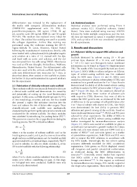
                     A                                         (Figure 1C). MSCs were seeded on scaffolds (or glass as
               PF n  s                                 (I)   control) and maintained in a growth medium for 3 days;
                     A
                      b                                        at the end, cells were stained with anti-CD90 (red) as a
            where  n is the number of enthesis scaffolds used in the   marker of MSCs [37]  and anti-actin (green) as a marker
            braiding process, and A  and A  are the cross-sections of the   of cytoskeleton organization. Both PLGA and PCL were
                              s
                                    b
            single enthesis scaffold specimen and the braided scaffold   able to support the MSC adhesion, in accordance with
            approximated with a cylindrical shape, respectively. The PF   the MTS results. Interestingly, the two solvent-casting
            is used to calculate the effective elastic modulus E  of the   polymers differently modified the MSC cytoskeleton
                                                    b
            3D braided scaffolds with the following relationship:  organization. Cells grown on PLGA displayed overall
                                                               cell  elongation  and  parallel  cytoskeletal  conformation,
                   E
               E =                                     (II)    as evidenced during the MSC differentiation to
                b  PF                                          tenocytes [38] . Conversely, several studies have reported
            where E is the elastic modulus calculated from the stress–  that MSCs exhibit a star shape during osteogenic
            strain curve without considering air gaps. At this stage,   lineage commitment [39,40] . In this respect, as evidenced
            no  biological  tests  were  conducted.  Scaffolds  were  only   by immunofluorescence images, PCL mainly supported
            mechanically characterized.                        star shape actin organization. These results prompted us


            Volume 9 Issue 5 (2023)                        301                         https://doi.org/10.18063/ijb.763

