
Page 35 - IJB-9-5
P. 35
International Journal of Bioprinting 3D bioprinted vascularized tissue models
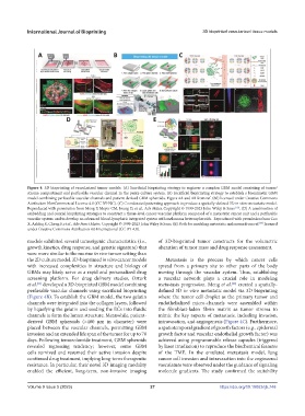
Figure 4. 3D bioprinting of vascularized tumor models. (A) Sacrificial bioprinting strategy to engineer a complex GBM model consisting of tumor/
stroma compartment and perfusable vascular channel in the penta-culture system. (B) Sacrificial bioprinting strategy to establish a biomimetic GBM
model combining perfusable vascular channels and patient-derived GBM spheroids. Figure 4A and 4B from ref. (60) licensed under Creative Commons
Attribution NonCommercial License 4.0 (CC BY-NC). (C) Coordinated patterning approach to produce a spatially-defined 3D in vitro metastatic model.
Reproduced with permission from Meng F, Meyer CM, Joung D, et al., Adv Mater, Copyright © 1999-2023 John Wiley & Sons . (D) A combination of
[62]
embedding and coaxial bioprinting strategies to construct a tissue-level cancer-vascular platform composed of a metastatic cancer unit and a perfusable
vascular system and to develop an advanced blood-lymphatic integrated system with melanoma heterospheroids. Reproduced with permission from Cao
X, Ashfaq R, Cheng F, et al., Adv Funct Mater, Copyright © 1999-2023 John Wiley & Sons. (E) Both for modeling metastatic melanoma from ref. licensed
[66]
under Creative Commons Attribution 4.0 International (CC BY 4.0).
models exhibited several tumorigenic characteristics (i.e., of 3D-bioprinted tumor constructs for the volumetric
growth kinetics, drug response, and genetic signature) that alteration of tumor mass and drug response assessment.
were more similar to the murine in vivo tumor setting than
the 2D culture model. 3D-bioprinted in vitro cancer models Metastasis is the process by which cancer cells
with increased complexities in structure and biology of spread from a primary site to other parts of the body
GBMs may likely serve as a rapid and personalized drug moving through the vascular system. Thus, establishing
screening platform. For drug delivery studies, Ozturk a vascular network plays a crucial role in modeling
et al. developed a 3D-bioprinted GBM model combining metastasis progression. Meng et al. created a spatially-
[61]
[62]
perfusable vascular channels using sacrificial bioprinting defined 3D in vitro metastatic model via 3D bioprinting
(Figure 4B). To establish the GBM model, the two gelatin where the tumor cell droplet as the primary tumor and
channels were integrated into the collagen layers, followed endothelialized micro-channels were assembled within
by liquefying the gelatin and seeding the ECs into fluidic the fibroblast-laden fibrin matrix as tumor stroma to
channels to form the lumen structure. Meanwhile, patient- mimic the key aspects of metastasis, including invasion,
derived GBM spheroids (>400 μm in diameter) were intravasation, and angiogenesis (Figure 4C). Furthermore,
placed between the vascular channels, permitting GBM a spatiotemporal gradient of growth factors (e.g., epidermal
invasion and an extended life span of the tumor for up to 70 growth factor and vascular endothelial growth factor) was
days. Following temozolomide treatment, GBM spheroids achieved using programmable release capsules (triggered
revealed regressing tendency; however, some GBM by laser irradiation) to reproduce the biochemical features
cells survived and resumed their active invasion despite of the TME. In the emulated metastasis model, lung
continued drug treatment, implying long-term therapeutic cancer cell invasion and intravasation into the engineered
resistance. In particular, their novel 3D imaging modality vasculature were observed under the guidance of signaling
enabled the efficient, long-term, non-invasive imaging molecule gradients. The study confirmed the suitability
Volume 9 Issue 5 (2023) 27 https://doi.org/10.18063/ijb.748