
Page 414 - IJB-9-5
P. 414
International Journal of Bioprinting 3D-printed Mg scaffolds promote bone defect repair
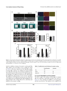
Figure 1. Characterization of magnesium (Mg) alloy coating samples. (A) The cross-sectional image of the coating using SEM. (B) EDS plane-scan results
of Mg alloy bulk samples. (C) Pictures of Mg alloy scaffolds. (D) SEM surface micrographs of Mg alloy scaffolds. (E) Pore size (left) and coating thickness
(right) of Mg alloy scaffolds. (F) Contact angle and sliding angle of Mg alloy samples to water (left) and hexadecane (right) before and after being coated
with polysilazane. *P < 0.05 and P < 0.05 vs. Mg group.
#
interfacial adhesion between the ceramic coating and Mg Table 2. Pencil hardness and adhesion of ceramic coating
alloy sample and the hardness of the coating are listed
in Table 2. The pencil hardness of the Mg alloy sample Pencil hardness Adhesion
increased after coating. The coating was closely bonded Mg 3H –
to the Mg alloy substrate, particularly in the Mg/Sc group. Mg/Sc 4H 5B
The adhesion grade of the coating in the Mg/Sc group was Mg/Sc/ZA 4H 4B
5B, and that of the Mg/SC/ZA group was 4B, which meets
the application requirements of most clinical scenarios. 3.2. In vitro degradation and drug release
The adhesion of the Mg/Sc/ZA group was lower than that The SEM images of the surface morphologies of samples
of the Mg/Sc group, possibly because, after loading ZA, the after the removal of degradation products are shown in
drug particles in the coating affected the uniformity and Figure 2A. The corrosion of scaffolds in the Mg group
integrity of the ceramic coating. was the most obvious. The structure of the scaffolds was
Volume 9 Issue 5 (2023) 406 https://doi.org/10.18063/ijb.769