
Page 419 - IJB-9-5
P. 419
International Journal of Bioprinting 3D-printed Mg scaffolds promote bone defect repair
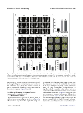
Figure 6. Radiological evaluation of osteoporotic bone defect healing. (A) Anterior and lateral X-ray images of distal femur of osteoporotic rats. (B)
Micro-CT of femoral samples of osteoporotic rats. (C) Quantification of residual volume percentage. (D) Three-dimensional reconstruction of implant.
(E) Quantification of the parameters included BV/TV, Tb.N, Tb.Sp, and BMD of BV. 3W, 6W, and 9W in (A), (B), and (D) represent 3 weeks, 6 weeks, and
9 weeks, respectively. *P < 0.05 vs. Mg group; P < 0.05 vs. Mg/Sc group.
#
the fluorescence intensity of reactive oxygen species (ROS) significantly higher than those in the Mg and Mg/Sc groups.
in the Mg/Sc/ZA group was significantly lower than that in The degradation rate of the 3D-printed Mg alloy scaffolds
the control and Mg/Sc groups. Cell survival was lower in in the Mg/Sc and Mg/Sc/ZA groups was also significantly
the Mg group, and the fluorescence intensity of ROS was the lower than that in the Mg group. The degradation of the
lowest in the Mg group (Figure 5A and B). Mg alloy scaffolds was similar to that induced by X-rays.
Quantitative analysis of the residual volume of the scaffolds
3.6. Effect of 3D-printed Mg alloy scaffolds on confirmed this observation (Figure 6C). Micro-CT scans
osteoporotic bone defect repair and reconstructed images of each group at different time
3.6.1. Radiographic analysis points are shown in Figure 6B and D. The results showed
The anteroposterior and lateral X-ray films of femur at that new bone tissue appeared around the 3D-printed Mg
different time points in each group are shown in Figure 6A. alloy porous scaffolds in the Mg/Sc/ZA group 3 weeks after
The defect healing rate in the Mg/Sc/ZA group was implantation, and a large amount of new bone tissue grew
Volume 9 Issue 5 (2023) 411 https://doi.org/10.18063/ijb.769