
Page 87 - IJB-9-5
P. 87
International Journal of Bioprinting Biocompatible BSA-GMA and TPP of 3D hydrogels with free radical type I photoinitiator
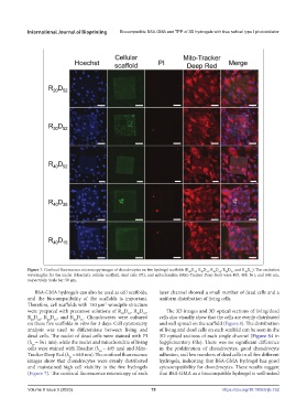
Figure 7. Confocal fluorescence microscopy images of chondrocytes on five hydrogel scaffolds (R D , R D , R D , R D , and R D ). The excitation
52
40
35
40
52
20
52
30
15
40
wavelengths for the nuclei (Hoechst), cellular scaffold, dead cells (PI), and mitochondria (Mito-Tracker Deep Red) were 405, 488, 561, and 640 nm,
respectively. Scale bar: 50 μm.
BSA-GMA hydrogels can also be used as cell scaffolds, laser channel showed a small number of dead cells and a
and the biocompatibility of the scaffolds is important. uniform distribution of living cells.
Therefore, cell scaffolds with 150 μm woodpile structure
2
were prepared with precursor solutions of R D , R D , The 3D images and 3D optical sections of living/dead
52
20
52
30
R D , R D , and R D . Chondrocytes were cultured cells also visually show that the cells are evenly distributed
15
40
40
52
35
40
on these five scaffolds in vitro for 3 days. Cell cytotoxicity and well spread on the scaffold (Figure 8). The distribution
analysis was used to differentiate between living and of living and dead cells on each scaffold can be seen in the
dead cells. The nuclei of dead cells were stained with PI 3D optical sections of each single channel (Figure S4 in
(λ = 561 nm), while the nuclei and mitochondria of living Supplementary File). There was no significant difference
ex
cells were stained with Hoechst (λ = 405 nm) and Mito- in the proliferation of chondrocytes, good chondrocyte
ex
Tracker Deep Red (λ = 640 nm). The confocal fluorescence adhesion, and low numbers of dead cells in all five different
ex
images show that chondrocytes were evenly distributed hydrogels, indicating that BSA-GMA hydrogel has good
and maintained high cell viability in the five hydrogels cytocompatibility for chondrocytes. These results suggest
(Figure 7). The confocal fluorescence microscopy of each that BSA-GMA as a biocompatible hydrogel is well-suited
Volume 9 Issue 5 (2023) 79 https://doi.org/10.18063/ijb.752