Page 256 - IJB-9-6
P. 256

International Journal of Bioprinting                           3D-Printed GelMA biomaterials in cartilage repair


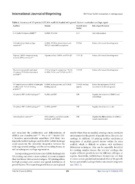
            Table 2. Summary of 3D-printed GELMA scaffolds loaded with growth factors in articular cartilage repair
             Scaffolds                  Bioinks                     Growth factor   Role of growth factors                          Characteristics of scaffolds    Results
                                                                    loaded
                                                                                                                                                                    In vitro                              In vivo
             IL-4-loaded bi-layer scaffolds [44]  GelMA, PCL-HA     IL-4          Anti-inflammation                                 •  Multi-layers with different bio-inks and   •  Both layers supported cell adhesion and proliferation  •  New cartilage and subchondral
                                                                                                                                      different functions           •  The upper layer relieved the inflammation of   bone regeneration
                                                                                                                                    •  Upper: GelMA; lower: PCL-HA    chondrocytes induced by IL-1b       •  Good mechanical strength similar
                                                                                                                                                                    •  The lower layer promoted osteogenesis  to that of native cartilage
             Cell-laden bioprinted cartilage   GelMA, PEGDA, photoinitiator, and   TGF-β1  Induce cells toward chondrogenesis       •  Fabricated via a core-shell electrospraying   •  Cells and nanospheres were evenly distributed  •  None
             construct [38]             TGF-β1-embedded nanospheres                                                                   technique                     •  Highest cell viability and proliferation on 5%/10%
                                                                                                                                    •  Modulus and swelling ratio could be adjusted   (PEGDA/GelMA) hydrogel
                                                                                                                                      by the addition of different PEGDA  •  Improved chondrogenic differentiation of
                                                                                                                                    •  Sustained release of TGF-β1    encapsulated MSCs.
             Alginate-GelMA interpenetrating   Alginate sulfate, GelMA, and TGF-β3  TGF- β3  Induce cells toward chondrogenesis     •  Maintained viscosity, shear-thinning and   •  Supported viability and robust chondrogenesis of   •  Supported chondrogenesis in vivo
             network (IPN) constructs [39]                                                                                            thixotropic properties          MSCs                                •  Controlled release of TGF-β3
                                                                                                                                    •  High-fidelity bioprinting                                            promoted cartilage-specific ECM
                                                                                                                                    •  Increased stiffness, and maintained resilience                       deposition
                                                                                                                                      and toughness
                                                                                                                                    •  Sustained release of TGF- β3
             Microenvironmentally optimized   A 3D printing ink containing D-ECM,   TGF-β3  Induce cells toward chondrogenesis      •  Microenvironment regulation  •  Directed endogenous stem/progenitor cell migration   •  Improved tissue repair outcomes in
             3D-printed TGF-β-functionalized   GelMA, PLGA, and TGF-β3, and PCL                                                                                       and differentiation                   the sheep animal model
             scaffolds [40]                                                                                                                                                                               •  Guided more organized neotissue
                                                                                                                                                                                                            formation
                                                                                                                                                                                                          •  Recapitulated the anisotropic
                                                                                                                                                                                                            structure
             3D-printed porous scaffolds of hydrogels  GelMA, hydroxyapatite, and TGF-β1-  TGF-β1-binding   Induce the endogenous TGF-β1   •  Multi-layers with different components and   •  Induced cartilage and osteogenic differentiation  •  Promoted osteochondral repair of
             modified with TGF-β1-binding   binding peptide         peptide       recruitment for chondrogenesis                      function                                                              rats
             peptides [41]                                                                                                          •  Upper: GelMA, TGF-β1 binding peptide;                              •  Recovered the animal gait behavior
                                                                                                                                      lower: GelMA, hydroxyapatite
             3D-printed PRP-GelMA hydrogels [49]  GelMA and PRP     PRP           Regulate the behaviors of BMSCs and               •  Fabricated using the digital micro-mirror   •  Promoted proliferation, migration, and osteogenesis   •  More cartilage and subchondral
                                                                                  macrophage                                          device (DMD) technique          and chondrogenesis of BMSCs by 20% PRP/GelMA  bone regeneration
                                                                                                                                                                    •  Promoted M2 polarization by 20% PRP/GelMA  •  More M2 macrophage infiltration
                                                                                                                                                                    •  Similar biological roles in BMSCs but less   and less M1 macrophage
                                                                                                                                                                      osteogenesis by 50% PRP/GelMA         presentation
             3D-printed PRP-GelMA hydrogels [50]  GelMA and PRP     PRP           Regulate the behaviors of cells                   •  Photoactivated PRP-based patient-specific   •  Long-term and constant rate growth factor release  •  Facilitated the proliferation and
                                                                                                                                      bioink                        •  Bioactivity protection of PRP        differentiation of the ATDC5 cells
                                                                                                                                    •  Had the desired mechanical properties (low   •  Satisfactory mechanical characteristics
                                                                                                                                      degradation rate and high mechanical strength)
             Osteochondral construct [51]  PRP, AdMSCs, and ECM mimetic   PRP     Regulate the differentiation of AdMSCs            •  Gradual printing of bio-inks  •  Induced glycosaminoglycan and calcium secretion,   •  None
                                        hydrogel, and GelMA                       toward chondrocytes                               •  Relatively low degradation rate and high   mineralization, and ECM production
                                                                                                                                      mechanical strength           •  Upregulated bone- and cartilage-unique genes
                                                                                                                                    •  Tissue-specific biomimetic structure


            and stimulates the proliferation and differentiation of   exactly where they are needed, creating a more conducive
            BMSCs into chondrocytes [35,36] . Wu  et  al.  loaded IGF-  environment for the growth of specific tissue, like articular
                                              [37]
            1 bioactive supramolecular nanofibers (IGF-1bsn) into   cartilage. In addition, 3D printing enables simultaneous
            injectable GelMA hydrogel, and this BSN-GelMA hydrogel   integration of multiple growth factors within the same
            could accelerate the interstitial integration between the   scaffold, which is difficult to achieve with traditional
            early regenerated cartilage and the surrounding tissues, as   fabrication techniques. This can be especially beneficial
            well as subsequent cartilage regeneration.         for creating complex tissues, like articular cartilage, that
               Incorporating growth factors into GelMA hydrogels for   require different growth factors for different cell types or
            3D printing can create a more optimal microenvironment   regions. Meanwhile, GelMA-based biomaterials can help
            than traditional fabrication techniques. 3D printing allows   to ensure a more gradual and sustained effect of the growth
            for high precision and control over spatial distribution of   factors, potentially leading to better outcomes in long-term
            growth factors. This means that growth factors can be placed   use (Table 2).

            Volume 9 Issue 6 (2023)                        248                         https://doi.org/10.36922/ijb.0116
   251   252   253   254   255   256   257   258   259   260   261