
Page 257 - IJB-9-6
P. 257
International Journal of Bioprinting 3D-Printed GelMA biomaterials in cartilage repair
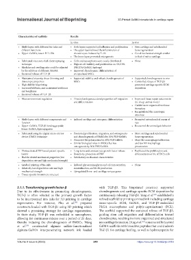
Table 2. Summary of 3D-printed GELMA scaffolds loaded with growth factors in articular cartilage repair
Scaffolds Bioinks Growth factor Role of growth factors Characteristics of scaffolds Results
loaded
In vitro In vivo
IL-4-loaded bi-layer scaffolds [44] GelMA, PCL-HA IL-4 Anti-inflammation • Multi-layers with different bio-inks and • Both layers supported cell adhesion and proliferation • New cartilage and subchondral
different functions • The upper layer relieved the inflammation of bone regeneration
• Upper: GelMA; lower: PCL-HA chondrocytes induced by IL-1b • Good mechanical strength similar
• The lower layer promoted osteogenesis to that of native cartilage
Cell-laden bioprinted cartilage GelMA, PEGDA, photoinitiator, and TGF-β1 Induce cells toward chondrogenesis • Fabricated via a core-shell electrospraying • Cells and nanospheres were evenly distributed • None
construct [38] TGF-β1-embedded nanospheres technique • Highest cell viability and proliferation on 5%/10%
• Modulus and swelling ratio could be adjusted (PEGDA/GelMA) hydrogel
by the addition of different PEGDA • Improved chondrogenic differentiation of
• Sustained release of TGF-β1 encapsulated MSCs.
Alginate-GelMA interpenetrating Alginate sulfate, GelMA, and TGF-β3 TGF- β3 Induce cells toward chondrogenesis • Maintained viscosity, shear-thinning and • Supported viability and robust chondrogenesis of • Supported chondrogenesis in vivo
network (IPN) constructs [39] thixotropic properties MSCs • Controlled release of TGF-β3
• High-fidelity bioprinting promoted cartilage-specific ECM
• Increased stiffness, and maintained resilience deposition
and toughness
• Sustained release of TGF- β3
Microenvironmentally optimized A 3D printing ink containing D-ECM, TGF-β3 Induce cells toward chondrogenesis • Microenvironment regulation • Directed endogenous stem/progenitor cell migration • Improved tissue repair outcomes in
3D-printed TGF-β-functionalized GelMA, PLGA, and TGF-β3, and PCL and differentiation the sheep animal model
scaffolds [40] • Guided more organized neotissue
formation
• Recapitulated the anisotropic
structure
3D-printed porous scaffolds of hydrogels GelMA, hydroxyapatite, and TGF-β1- TGF-β1-binding Induce the endogenous TGF-β1 • Multi-layers with different components and • Induced cartilage and osteogenic differentiation • Promoted osteochondral repair of
modified with TGF-β1-binding binding peptide peptide recruitment for chondrogenesis function rats
peptides [41] • Upper: GelMA, TGF-β1 binding peptide; • Recovered the animal gait behavior
lower: GelMA, hydroxyapatite
3D-printed PRP-GelMA hydrogels [49] GelMA and PRP PRP Regulate the behaviors of BMSCs and • Fabricated using the digital micro-mirror • Promoted proliferation, migration, and osteogenesis • More cartilage and subchondral
macrophage device (DMD) technique and chondrogenesis of BMSCs by 20% PRP/GelMA bone regeneration
• Promoted M2 polarization by 20% PRP/GelMA • More M2 macrophage infiltration
• Similar biological roles in BMSCs but less and less M1 macrophage
osteogenesis by 50% PRP/GelMA presentation
3D-printed PRP-GelMA hydrogels [50] GelMA and PRP PRP Regulate the behaviors of cells • Photoactivated PRP-based patient-specific • Long-term and constant rate growth factor release • Facilitated the proliferation and
bioink • Bioactivity protection of PRP differentiation of the ATDC5 cells
• Had the desired mechanical properties (low • Satisfactory mechanical characteristics
degradation rate and high mechanical strength)
Osteochondral construct [51] PRP, AdMSCs, and ECM mimetic PRP Regulate the differentiation of AdMSCs • Gradual printing of bio-inks • Induced glycosaminoglycan and calcium secretion, • None
hydrogel, and GelMA toward chondrocytes • Relatively low degradation rate and high mineralization, and ECM production
mechanical strength • Upregulated bone- and cartilage-unique genes
• Tissue-specific biomimetic structure
5.1.1. Transforming growth factor-β with TGF-β3. This bioprinted construct supported
Due to its effectiveness in promoting chondrogenesis, chondrogenesis and cartilage-specific ECM deposition by
[40]
TGF-β is often selected as the primary growth factor continuously releasing TGF-β3. Yang et al. established a
to be incorporated into inks for 3D printing in cartilage refined scaffold by printing a mixed ink including cartilage
regeneration. For instance, Zhu et al. prepared tissue-specific ECM, GelMA, and TGF-β3-embedded
[38]
constructs loaded with TGF-β1 using 3D printing which PLGA microspheres and poly(ε-caprolactone) (PCL).
showed a promising strategy for cartilage regeneration. The scaffold supported the sustained release of TGF-β3,
In their study, TGF-β1 was embedded in nanospheres, guiding stem cell migration and differentiation toward
allowing for continuous release over a period of 21 days, chondrocytes, resulting in more organized and structured
[41]
thereby inducing the chondrogenesis of BMSCs. Wang neocartilage formation. Ding et al. constructed bi-layered
et al. constructed alginate sulfate-functionalized GelMA scaffolds with bioactive peptides that could adsorb
[39]
alginate-GelMA interpenetrating network ink loaded TGF-β1 for cartilage healing, as well as hydroxyapatite for
Volume 9 Issue 6 (2023) 249 https://doi.org/10.36922/ijb.0116