
Page 268 - IJB-9-6
P. 268
International Journal of Bioprinting 3D bioprinting for vascular system
[24]
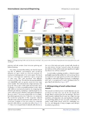
Figure 1. (A) Design drawing of valve multi-level structure modeling . (B) An aortic valve mold made of sugar glass and a time-lapse photo of the mold
[26]
dissolving .
printing methods contain direct extrusion printing and the root of the valve and aortic stromal cells directly in
indirect printing. the valve lobules. On day 7 of graft culture, the strength
First, direct extrusion bioprinting can directly deposit and stiffness of the cell material were significantly higher
[25]
bio-inks of different concentrations and containing than that of the cell-free scaffold .
different cell types, which can effectively replicate the Second, indirect printing provides a relatively simple
mechanical heterogeneity of the valve region. The heart and highly reproducible method for valve printing. Rioux
valve is a heterogeneous anatomical region composed et al. used low-cost sugar glass to print the stent mold and
of valve ring, valve, and notochord with different then filled it with sodium alginate hydrogel, crosslinking it
mechanical properties. The valve has strong expandability with CaCl . A complete aortic valve is produced after the
2
and adaptability and can quickly open and close large glass-lined mold dissolves (Figure 1B) .
[26]
blood vessels. The notochord has a high hardness that
keeps the lumen open under harsh hemodynamic load. 3. 3D bioprinting of small-caliber blood
Hockaday et al. have successfully produced aortic valves
with different mechanical properties by designing two vessels
different gel schemes, including a rigid hydrogel for the The research and development of small-diameter vascular
aortic notochord and a flexible, expandable hydrogel for prostheses has been a hot topic in the past decade, but
the aortic valve . The cross-section of the valve is a multi- formal clinical products have not yet emerged. The ideal
[23]
layer heterogeneous structure. Vukicevic et al. compared small-diameter artificial blood vessel has a high forming
the mechanical properties of the composite material resolution, cell integration rate, and mechanical strength.
with the target natural components and constructed the 3D bioprinting allows for the successful construction of
multi-layer structure of the valve using the composite perfect small-caliber blood vessels by combining new
material with different concentration ratios (Figure 1A) bio-inks, well-designed printing strategies, and multiple
[24] . Duan et al. wrapped smooth muscle cells directly in manufacturing methods.
Volume 9 Issue 6 (2023) 260 https://doi.org/10.36922/ijb.0012