
Page 270 - IJB-9-6
P. 270
International Journal of Bioprinting 3D bioprinting for vascular system
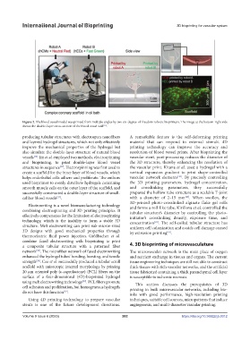
Figure 2. The blood vessel model was printed from multiple angles by two six-degree-of-freedom robotic bioprinters. The image at the bottom-right side
shows the double-layer cross-section of the blood vessel wall .
[35]
producing tubular structures with electrospun nanofibers A remarkable feature is the self-deforming printing
and layered hydrogel structures, which not only effectively material that can respond to external stimuli. 4D
improve the mechanical properties of the hydrogel but printing technology can improve the accuracy and
also simulate the double-layer structure of natural blood resolution of blood vessel prints. After bioprinting the
vessels . Jin et al. employed two methods, electrospinning vascular stent, post-processing reduces the diameter of
[36]
and bioprinting, to print double-layer blood vessel the 3D structure, thereby enhancing the resolution of
structures in sequence . Electrospinning was first used to the vascular print. Kitana et al. used a hydrogel with a
[37]
create a scaffold for the inner layer of blood vessels, which vertical expansion gradient to print shape-controlled
helps endothelial cells adhere and proliferate. The authors vascular network elements [40] . By precisely controlling
used bioprinter to evenly distribute hydrogels containing the 3D printing parameters, hydrogel concentration,
smooth muscle cells on the outer layer of the scaffold, and and crosslinking parameters, they successfully
successfully constructed a double-layer structure of small- prepared the hollow tube structure as a scalable T-joint
caliber blood vessels . with a diameter of 2–15 mm [40] . When swollen, the
[37]
3D-printed photo-crosslinked alginate flake gel rolls
Electrowriting is a novel biomanufacturing technology
combining electrospinning and 3D printing principles. It and forms a roll-like tube. Kirillova et al. controlled the
tubular structure’s diameter by controlling the photo-
effectively compensates for the limitation of electrospinning initiator’s crosslinking density, exposure time, and
technology, which is the inability to form a stable 3D concentration [41] . The self-coiled tubular structure has
structure. Melt electrowriting can print sub-micron-sized uniform cell colonization and avoids cell damage caused
3D designs with good mechanical properties through by extrusion printing [41] .
thermoelectric fluid power injection. Größbacher et al.
combine fused electrowriting with bioprinting to print
a composite tubular structure with a patterned fiber 4. 3D bioprinting of microvasculature
network . The microfiber network of fused electrowriting The microvascular network is the main place of oxygen
[38]
enhanced the hydrogel tubes’ bending, bursting, and tensile and nutrient exchange in tissues and organs. The current
[38]
strength . Cao et al. successfully produced a tubular scroll tissue engineering techniques are still not able to construct
scaffold with anisotropic internal morphology by printing thick tissues with rich vascular networks, and the artificial
20 μm oriented poly (ε-caprolactone) (PCL) fibers on the tissue fabricated containing a thick parenchymal cell layer
surface of a four-dimensional (4D)-bioprinted hydrogel is susceptible to ischemic necrosis.
using melt electrowriting technology . PCL fibers promote This section discusses the prerequisites of 3D
[39]
cell adhesion and proliferation, but homogeneous hydrogels printing in built microvascular networks, including bio-
do not have this function .
[39]
inks with good performance, high-resolution printing
Using 4D printing technology to prepare vascular techniques, suitable cell sources, micropatterns that induce
stents is one of the future development directions. angiogenesis, and multi-diameter vascular printing.
Volume 9 Issue 6 (2023) 262 https://doi.org/10.36922/ijb.0012