
Page 275 - IJB-9-6
P. 275
International Journal of Bioprinting 3D bioprinting for vascular system
[69]
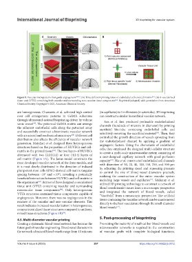
Figure 5. Vascular micropatterns that guide angiogenesis [64,66] . (A) Three different printing ratios of endothelial cells and cell-matrix . (B) A vascularized
[46]
tissue unit (VTU) containing both vascular and surrounding non-vascular tissue components . Reprinted (adapted) with permission from American
Chemical Society. Copyright © 2021, American Chemical Society.
are homogeneous. Chansoria et al. achieved high control (in capillaries) to 5 millimeters (in arterioles). 3D bioprinting
over cell arrangement patterns in GelMA substrates can construct a similar hierarchical vascular network.
through ultrasound-assisted bioprinting driven by volume Son et al. first produced perfusable endothelialized
sonic waves . The patterned GelMA matrix can arrange channels (hundreds of microns in diameter) by printing
[68]
the adherent endothelial cells along the patterned array sacrificial bio-inks containing endothelial cells and
and successfully construct a biomimetic vascular network selectively removing the sacrificial material . Then, they
[70]
with structural and mechanical anisotropy . Different cell controlled the growth direction of vessels sprouting from
[68]
distribution also affects the efficiency of vascular network the endothelialized channel by applying a gradient of
generation. Maiullari et al. designed three heterogeneous angiogenic factors. Using the chemotaxis of endothelial
structures based on the proportion of HUVECs and cell- cells, they employed the designed multi-cellular structure
matrix in the printed tissue . The two layers of HUVECs to create a multi-scale microvascular system consisting of
[69]
alternated with two (2:2:2:2:2) or four (4:2:4) layers of a user-designed capillary network with good perfusion
cell-matrix (Figure 5A). The Janus model constructs the capacity . Nie et al. constructed endothelial cell channels
[70]
most developed vascular network of the three models, and with diameters of 10, 20, 40, 100, 150, 250, and 500 μm
it is most clearly distributed in the direction of induced by adjusting the printing speed and squeezing pressure
pluripotent stem cells (iPSC)-derived cell-matrix (angular to control the size of blood vessel diameters precisely,
spacing between -10° and +10°), revealing a potentially realizing the construction of the entire vascular system
beneficial interaction between HUVECs and cell-matrix in including large vessels and capillaries . Szklanny et al.
[71]
the organization . Barrs et al. have designed a vascularized utilized 3D printing technology to construct a network of
[69]
tissue unit (VTU) containing vascular and surrounding blood vessels inside tissues from a microscopic perspective
nonvascular tissue components . Only heterogeneous and integrated the network of blood vessels, called
[46]
VTUs structures contained tissue components capable of “VascFold,” from a mesoscopic perspective. The artificial
angiogenesis. Moreover, there was no angiogenesis in the tissue containing the vascular network can be anastomosed
mixture of the vascular and non-vascular elements. This directly to the host vasculature through the small-diameter
result indicates increased vascularization in heterogeneous, blood vessels .
[72]
compartmentalized tissue structures compared to uniform,
mixed tissue structures (Figure 5B) .
[46]
5. Post-processing of bioprinting
4.5. Multi-diameter vascular printing
Creating a systematic blood vessel network has become the Promoting the maturity of small-caliber blood vessels and
future goal of vascular engineering. Blood vessel diameters in microvascular networks is required in the construction
the network of natural blood vessels range from 10 microns of vascular grafts with complete biological functions.
Volume 9 Issue 6 (2023) 267 https://doi.org/10.36922/ijb.0012