
Page 272 - IJB-9-6
P. 272
International Journal of Bioprinting 3D bioprinting for vascular system
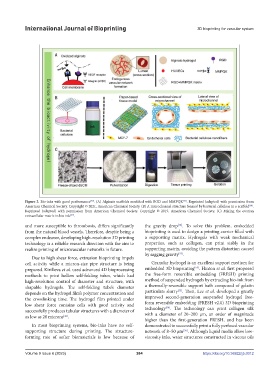
Figure 3. Bio-inks with good performance . (A) Alginate scaffolds modified with RGD and MMPQK . Reprinted (adapted) with permission from
[46]
[40]
American Chemical Society. Copyright © 2021, American Chemical Society. (B) A microchannel structure formed by bacterial cellulose in a scaffold .
[48]
Reprinted (adapted) with permission from American Chemical Society. Copyright © 2019, American Chemical Society. (C) Mixing the ovarian
extracellular matrix in bio-ink .
[49]
and more susceptible to thrombosis, differs significantly the gravity drop . To solve this problem, embedded
[12]
from the natural blood vessels. Therefore, despite being a bioprinting is used to design a printing carrier filled with
complex endeavor, developing high-resolution 3D printing a supporting matrix. Hydrogels with weak mechanical
technology is a reliable research direction with the aim to properties, such as collagen, can print stably in the
realize printing of microvascular networks in future. supporting matrix, avoiding the pattern distortion caused
by sagging gravity .
[53]
Due to high shear force, extrusion bioprinting impels
cell activity while a micron-size pipe structure is being Granular hydrogel is an excellent support medium for
[54]
prepared. Kirillova et al. used advanced 4D bioprocessing embedded 3D bioprinting . Hinton et al. first proposed
methods to print hollow self-folding tubes, which had the free-form reversible embedding (FRESH) printing
high-resolution control of diameter and structure, with method of suspended hydrogels by extruding bio-ink from
shapable hydrogels. The self-folding tube’s diameter a thermally-reversible support bath composed of gelatin
[55]
depends on the hydrogel film’s polymer concentration and particulate slurry . Then, Lee et al. developed a greatly
the crosslinking time. The hydrogel film printed under improved second-generation suspended hydrogel free-
low shear force contains cells with good activity and form reversible embedding (FRESH v2.0) 3D bioprinting
[56]
successfully produces tubular structures with a diameter of technology . The technology can print collagen silk
as low as 20 microns . with a diameter of 20–200 µm, an order of magnitude
[41]
higher than the first-generation FRESH, and has been
In most bioprinting systems, bio-inks have no self- demonstrated to successfully print a fully perfused vascular
supporting structure during printing. The structure- network of 8–50 μm . Although liquid media allow low-
[56]
forming rate of softer biomaterials is low because of viscosity inks, water structures constructed in viscous oils
Volume 9 Issue 6 (2023) 264 https://doi.org/10.36922/ijb.0012