Page 310 - IJB-9-6
P. 310

International Journal of Bioprinting                                Progress in bioprinted ear reconstruction


            Table 2. Continued
             Study       Aim of study        Study   Animal   Study focus  3D printing   Components  Printed                     Printed   Cell nature/type  Notable post-  Assessment   Findings                    Limitations and suggested
                                             setting  model (if        technique                   shape                         material                printing       of success/                                  improvements
                                                   any)                                                                                                  modifications  integration
                                                                                                                                                                                    •  At 12 months, the reconstructed auricle   •  Multiple additional
                                                                                                                                                                                      presented high stiffness and low flexibility,   surgical steps
                                                                                                                                                                                      whereas at 24 months, an obvious   incorporated:
                                                                                                                                                                                      improvement in inflexibility with more   •  Tissue expanded
                                                                                                                                                                                      distinct structures were achieved.  preoperatively for 3
                                                                                                                                                                                    •  Among the total five cases, four cases showed   months (psychosocial
                                                                                                                                                                                      obvious cartilage formation after 6 months   impact)
                                                                                                                                                                                      post-implantation (one case was lost to   •  Split-thickness skin
                                                                                                                                                                                      follow-up).                        graft from groin was
                                                                                                                                                                                    •  MRI conformed a significant portion of PCL   required
                                                                                                                                                                                      has degraded (complete degradation of PCL   •  Scar revision
                                                                                                                                                                                      in vivo normally requires 2–4 years). Biopsied   surgeries were
                                                                                                                                                                                      samples revealed formation of mature in vivo   required at 6 and 18
                                                                                                                                                                                      cartilage at 6 months and 18 months post-  months
                                                                                                                                                                                      operatively.
            Abbreviations: ACM, ; ACMMA, methacrylate-modified acellular cartilage matrix; ASCs, adipose-derived stem cells; AuCPCs, auricular cartilage pro-
            genitor cells; CAD, computer-aided design; CAM, computer-aided manufacturing; CPS, cell-printed structure; CSHS, cell-seeded hybrid scaffold; CSS,
            cell-seeded scaffold; CT, computed tomography; DLP, digital light processing; DMEM, Dulbecco’s Modified Eagle Medium; ECM, extracellular matrix;
            FDM, fused deposition modeling; GAG, glycosaminoglycan; GelMA, gelatin methacrylate; H&E, hematoxylin and eosin staining; HA, hyaluronic acid;
            HAMA, hyaluronic acid methacrylate; MRI, magnetic resonance imaging; MSCs, mesenchymal stem cells; PBS, phosphate-buffered saline; PCL, poly-
            caprolactone; PEG, polyethylene glycol; PEO, poly(ethylene oxide); PGA, polyglycolic acid; PGLA, poly(lactic-co-glycolic acid); PLA, polylactic acid; PPU,
            perforated polyurethane; PRP, platelet-rich plasma; PU, polyurethane; SEM, scanning electron microscopy; SLS, selective laser sintering; UV, ultraviolet.

                                                               comparing scaffold materials and cartilage formation was
                                                               not possible.
                                                               4. Discussion

                                                               4.1. Overcoming current obstacles to clinical
                                                               translation
                                                               Auricular reconstruction is a challenging endeavor, partly
                                                               due to the complex 3D geometry of the auricle , so unique
                                                                                                   [4]
                                                               to each individual that the pinna has been proposed as a
                                                               forensic identifier .
                                                                             [5]
                                                                  This review demonstrates that 3D printing has the
                                                               potential to have a significant impact on this relatively
                                                               niche but complex area of reconstructive surgery by
                                                               enhancing current surgical reconstructive options, and
                                                               before clinical translation can truly occur, this technology
                                                               requires optimization in  multiple areas  and large-scale
                                                               clinical trials.
                                                                  The printed auricle’s desired anatomically detailed
                                                               shape should be ensured for a lifetime, even after
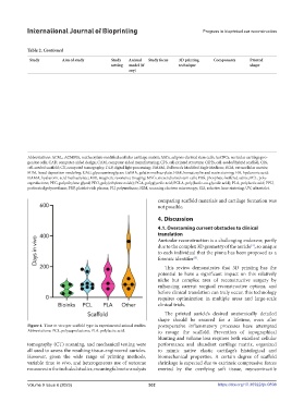
            Figure 4. Time in vivo per scaffold type in experimental animal studies.   postoperative inflammatory processes have attempted
            Abbreviations: PCL, polycaprolactone; PLA, polylactic acid.  to ravage the scaffold. Prevention of topographical
                                                               blunting and volume loss requires both excellent cellular
            tomography (CT) scanning, and mechanical testing were   performance and abundant cartilage matrix, organized
            all used to assess the resulting tissue-engineered auricles.   to mimic native elastic cartilage’s histological and
            However, given the wide range of printing methods,   biomechanical properties.  A certain  degree  of scaffold
            variable time in vivo, and heterogeneous use of outcome   shrinkage is expected due to extrinsic compressive forces
            measures in the included studies, meaningful meta-analysis   exerted  by the overlying  soft  tissue, myocontractile


            Volume 9 Issue 6 (2023)                        302                        https://doi.org/10.36922/ijb.0898
   305   306   307   308   309   310   311   312   313   314   315