
Page 362 - IJB-9-6
P. 362
International Journal of Bioprinting Surface modification of PCL scaffolds
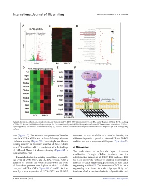
Figure 6. Surface modification mediated osteogenesis via integrinα2/β1-PI3K-AKT signaling pathway. (A) The scatter diagram of DEGs. (B) The heatmap
of DEGs. (C) The top 20 KEGG signaling pathways. (D) The expression of genes in PI3K-Akt signaling pathway. (E) The expression of proteins in PI3K-Akt
signaling pathway was detected by Western blotting. (F) Modified surface could facilitate osteogenic differentiation via integrinα2/β1-PI3K-Akt signaling
pathway.
sites (Figure 7G). Furthermore, the presence of lamellar decreased in both scaffolds at 3 months. Besides, the
bone in M-PCL scaffolds was confirmed through Masson’s difference in protein expression between PCL and M-PCL
trichrome staining (Figure 7H). Interestingly, van Gieson scaffolds was less pronounced at this point (Figure 8A–F).
staining revealed an increased number of bone calluses
in M-PCL scaffolds, which is consistent with the findings 4. Discussion
of H&E and Masson’s trichrome staining (Figure S2 in This study aimed to explore the impact of surface
Supplementary File). modification through alkaline treatment on the
Immunohistochemical staining was utilized to quantify osteoinductive properties of MEW PCL scaffolds. PCL
the levels of OPN, OCN, and RUNX2 protein. After a has been extensively utilized for creating biocompatible
duration of 1 month, the results indicated that the levels scaffolds for tissue engineering, particularly for bone tissue
of these three proteins were higher in M-PCL scaffolds engineering scaffolds . The limitations of PCL in tissue
[46]
compared to PCL scaffolds (Figure 8A, C, and E). As time engineering arise from its surface hydrophobicity and
went by, protein expression of OPN, OCN, and RUNX2 inertness, which are not conducive to cell proliferation and
Volume 9 Issue 6 (2023) 354 https://doi.org/10.36922/ijb.1071