
Page 363 - IJB-9-6
P. 363
International Journal of Bioprinting Surface modification of PCL scaffolds
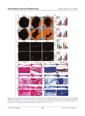
Figure 7. Micro-CT analysis, sequential fluorescent labeling, and histological analysis of new bone formation. (A) Micro-CT 3D images of PCL scaffolds.
(B) Quantitative analysis of BV/TV value after implantation for 1 and 3 months. (C, D) Quantitative analysis of bone formation according to Tb.Th and
Tb.Sp. (E) The newly formed bone was stained using calcein-Alizarin Red. (F) Quantitative analysis of mineral apposition rate. (G) H&E staining of the
decalcified sections. (H) Masson’s trichrome staining of the decalcified sections. *P < 0.05, **P < 0.01.
Volume 9 Issue 6 (2023) 355 https://doi.org/10.36922/ijb.1071