
Page 383 - IJB-9-6
P. 383
International Journal of Bioprinting Biomimetic biofabrication of tumors volume
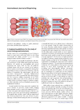
Figure 1. Tumor microenvironment (TME). The schematics summarize the principal cellular components of the TME with associated functions and the
support for the maintenance, progression, and ultimate treatment of the cancerous tissue.
associated macrophages, aiming to guide phenotype to resemble the TME, cancer cells are prone to adaptations
polarization and thus cancer regression. to in vitro growth, losing the native characteristics of
the tumor. Recently, patient-derived xenografts (PDX)
3. Engineering platforms for the study of have been frequently used since these models are able to
[36]
cancer biology and metastasis recapitulate the main characteristics of the host tumor .
On the other hand, animal models are more expensive
To date, most of our understanding of cancer biology is compared to 2D models, demanding, time-consuming, and
based on experiments performed either in vitro using more importantly, unable to mimic the actual response of
conventional 2D culture models or in vivo using a human organism . Furthermore, the high demand for
[31]
[37]
xenograft animal models of human tumors . test subjects needed for animal experimentation has raised
[32]
2D models are economically favorable due to the low- ethical concerns and has led to the founding of several
cost maintenance of the culture and related experiments. organizations demanding the replacement and reduction
[38]
However, 2D models are oversimplified and cannot of animals in research . To overcome the aforementioned
recapitulate the native TME. In fact, 2D platforms limitations, 3D culture platforms have gained increasing
comprise cancer cells that are cultured on flat surfaces, interest by more closely mimicking the TME and
forced to grow as monolayers, consequently altering their providing more physiologically relevant information.
morphology as well as several of their functions, critically Indeed, unlike cells cultured in plastic, 3D culture models
failing to recapitulate the physiological cell–cell and are not constrained to a single layer and the additional
cell–matrix interactions . Currently, bidimensional dimensionality allows for the spatial arrangement of their
[33]
platforms are not able to represent the tumor heterogeneity surface receptors and also induces physical constraints
[39]
and the numerous cellular components of the TME. among surrounding cells . In the attempt to create 3D
Moreover, unlike cancer cells in vivo, cells in 2D culture platforms that may recapitulate the pathophysiological
models receive a continuous supply of nutrients, oxygen, functionality of TME, 3D cancer models have been
and other molecules which are abundantly present in the developed, ranging from spheroids cultures to biomaterial
supplemented medium. scaffolds and tumor-on-a-chip platforms.
Animal models have been extensively employed for Multi-cellular tumor spheroids are a scaffold-free 3D
cancer research. In particular, mouse models, ranging cancer cell-only platform typically consisting of multiple
from xenograft tumors to genetically engineered mice , type of cell aggregates that favor cell–cell interactions and
[34]
are the most used model systems because of the low cost, produce their own ECM. As solid tumors, multi-cellular
small size, ease of use, and known genetic information . tumor spheroids (MCTS) display similar stiffness and
[35]
In xenograft tumor models, cancer cells are transplanted spatial heterogeneity and also similar nutrients, oxygen,
[40,41]
into immunocompromised mice and allowed to grow. and cell proliferation gradients . Even though more
Although cell line-derived xenografts have the advantage advanced compared to 2D culture models, a limiting
factor of MCTS is the lack of the actual range of ECM
Volume 9 Issue 6 (2023) 375 https://doi.org/10.36922/ijb.1022