
Page 113 - v11i4
P. 113
International Journal of Bioprinting 3D bioprinting for translational toxicology
robust, porous scaffolds, as shown in Figure 2I—further and adjustable stiffness. Composite formulations—such
96
expand the design space. as gelatin–alginate blends—have demonstrated enhanced
The selection of 3D bioprinting modalities requires mechanical integrity and cell compatibility for soft-tissue
97
meticulous evaluation of factors including cell viability, constructs. Non-animal polysaccharides like alginate and
structural complexity, and industrial scalability. Table 1 hyaluronic acid offer mild ionic or photo-crosslinking,
summarizes critical parameters for each modality, customizable mechanics, and minimal immunogenicity.
+
highlighting their proficiency in constructing multi-scale Alginate rapidly gels in divalent-cation baths (e.g., Ca² ),
toxicological models, spanning from micro-level precision supporting modular constructs and even laser-induced
98
to macro-level biomimicry. bioprinting with high cell viability. Silk-based inks—from
fibroin or recombinant spider silk—provide superior tensile
3.2. Design of bioinks: natural and strength and biocompatibility, though natural spider silk’s
99
synthetic materials scalability is limited. 100,101 Recombinant variants and silk-
In the realm of bioprinting technology, the choice of like polypeptides now enable tunable hydrogels and porous
bioink—the printing material—is crucial for the successful scaffolds for diverse tissue engineering applications.
99
construction of target structures. Commonly used bioinks dECM bioinks preserve organ-specific biochemical cues
primarily consist of natural and synthetic polymers, each (collagens, glycosaminoglycans, laminin) that direct cell
possessing distinct advantages and limitations. Synthetic phenotype and function. 102,103 For instance, cardiac and
materials excel in mechanical properties, rendering them cartilage dECM enhance cardiomyocyte maturation and
suitable for fabricating structures where lower biological upregulate SOX9/COL2A1 expression, respectively. Yet,
103
affinity is acceptable. Conversely, natural materials offer complex decellularization, variable gelation kinetics, and
superior biocompatibility and bioactivity but often exhibit insufficient native mechanics often necessitate chemical
weaker mechanical performance, making them well-suited crosslinkers or synthetic blends to balance printability
for mimicking native tissues. Therefore, the development with physiological performance. In summary, natural
102
and selection of appropriate bioinks are crucial for materials are highly valued in bioprinting for their
achieving the ultimate goals of bioprinting. excellent biocompatibility and bioactivity. However,
94
their mechanical limitations often require reinforcement
3.2.1. Natural materials through composite formulations. By carefully selecting and
Natural bioinks derive from biodegradable polymers that modifying natural materials, their potential applications in
hydrolyze or undergo enzymatic degradation in vivo, bioprinting can be substantially expanded.
yielding biocompatible byproducts. They are broadly
classified as animal-derived or non-animal-derived 3.2.2. Synthetic materials
materials. Among animal-derived polymers, collagen Synthetic polymers have precise control over mechanical
95
and its thermo-responsive derivative gelatin excel at properties, degradation rates, and batch-to-batch
recapitulating native ECM architecture. However, gelatin’s consistency, making them staples in bioprinting.
slow, temperature-dependent gelation compromises Polyethylene glycol hydrogels are Food and Drug
printing fidelity; methacrylation overcomes this by Administration-approved, highly hydrophilic, and
enabling rapid, tunable ultraviolet-mediated crosslinking resistant to protein fouling. Low-molecular-weight
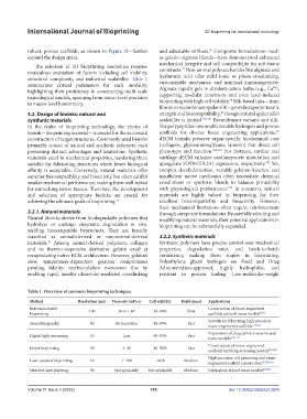
Table 1. Overview of common bioprinting techniques
Method Resolution (μm) Viscosity (mPa·s) Cell viability Build speed Applications
Extrusion-based 100 30–6 × 10 7 50–90% Slow Construction of tissue-engineered
bioprinting scaffolds and soft tissue models 76–79
Suitable for fabricating high-precision
Stereolithography 50 No limitation 85–95% Fast
tissue-engineered scaffolds 77,80–82
Preparation of drug delivery systems and
Digital light processing 10 Low 85–95% Fast
tissue models 81,83–86
Construction of tissue-engineered
Inkjet bioprinting 50 3–30 80–90% Fast
scaffolds and drug-screening models 84,87,88
High-precision cell patterning and tissue-
Laser-assisted bioprinting 10 1–300 ~90% Medium
engineered scaffold construction 75,77,89–91
Selective laser sintering 50 Not applicable Not applicable Medium Fabrication of hard tissue models 83,92,93
Volume 11 Issue 4 (2025) 105 doi: 10.36922/IJB025210209